
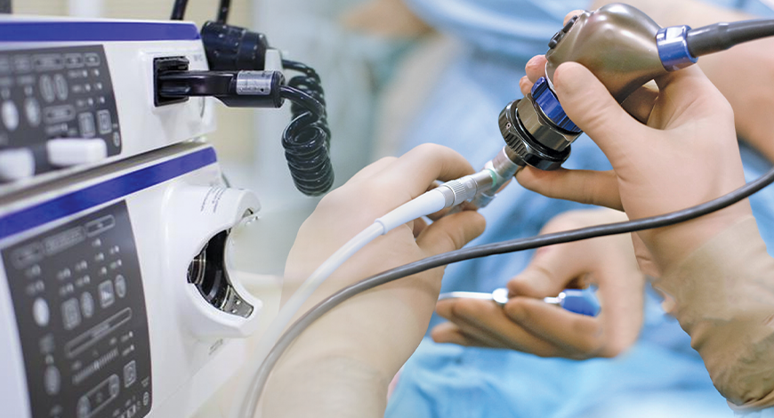
Endoscopy Equipment
A transformative AI journey ahead for endoscopy
Endoscopy will continue to thrive as a screening and diagnostic tool, aided by new technologies like AI. New technologies will improve endoscopy capabilities rather than replace them.

Over the next 10 years, gastrointestinal (GI) will evolve into a field that is increasingly specialized and patient-centered, driven by advances in technology.
With respect to increased specialization, the frontier of advanced endoscopy is already pushing the boundaries of what was previously only possible with surgery. This trend will continue with advanced endoscopy providing a more frequent and less invasive alternative to surgical management of disease. This trend is aided by advancements in endoscopic capabilities and other related tools.
Whereas disruptive technologies, particularly genetic and molecular-based testing, may make a dent in routine endoscopic procedures, newer procedures and technologies, like motorized enteroscopy, non-endoscopic diagnostic tests, will become available to fill the gap for general GIs.
For the therapeutic endoscopist, advanced resection platforms and robotic endoscopy would have come of age and be firmly established by 2030, thereby transforming third-space endoscopy and the entire endo-surgery field.
Endoscopic ultrasound interventions will continue to expand and bridge the gap between surgery and endoscopy. Defect-closure and bleeding-control devices will evolve to help conquer more complex endoscopic challenges, and will be simpler to use in emergency situations. Disposable endoscopes will have found several niche applications, especially in high-risk procedures and clinical scenarios. AI platforms would have matured by 2030, with specific applications aimed to improve yield of endoscopy and diagnostic testing.
Bariatric endoscopic practice will have evolved into more of a metabolic endoscopy approach, from mechanical to increasingly physiologically oriented interventions. Personalized cancer medicine and immunotherapy will be the norm for GI-cancer management. Similarly, immune-modulators and biologics will continue to transform IBD care, particularly for severe patients. Increased global connectivity and networking will result in greater opportunities for teaching, training, and research collaboration worldwide.
Ironically, technology will also replace some of our basic diagnostic GI procedures with noninvasive options. Further development of the intelligent capabilities of capsule endoscopy and similar ingestible diagnostics, and the development of novel biomarkers will likely supplant the need to use endoscopy as a primary form of diagnostic assessment. This will have broad implications for CRC screening as well as practice patterns in procedure-heavy practices.
Ten years from now, the world and the GI space will be quite different. Those who can adapt and evolve accordingly will be most successful. Despite all the changes and advancements, ultimate goal will and always should be to provide the highest-quality patient care possible.
Market trends
Endoscopy will continue to thrive as a screening and diagnostic tool, aided by new technologies like AI. New technologies will improve endoscopy capabilities rather than replace them. On a day-to-day basis, however, by 2030 or sooner, GIs will see their workflow and revenue sources evolve from traditional fee-for-service and endoscopy-focused workt to more of a fee-for-value and chronic disease management type of practice.
Consolidation will continue among payers and providers, which will continue to impact every aspect of healthcare. However, as healthcare services transition to more risk-based compensation models, larger entities will pursue broader strategic partnerships in an effort to manage risk. This represents an opportunity for large independent physician practices who will be better equipped to aggregate data and provide services to a broader market, with lower cost and greater efficiency.Endoscopic procedures will remain the cornerstone of the GI practice. Despite alternative CRC screening options, population demographics and a fixed number of physician trainees will result in continued high demand for endoscopic procedures. GI procedure volumes will continue to migrate away from the hospital outpatient departments to fully integrated and lower-cost ASCs. High demand for GI services may lead to challenges in physician recruitment.
Digital health applications and nanotechnologies are progressing at a rapid speed, and could enhance the way we treat and manage many GI diseases. The combination of sensor-based technology, via ingestion of nanoparticles for chemotherapeutic drug delivery and monitoring of levels, to genomic evaluation of the individual patient’s gut microbiome, will transform GI care as we know it. We will become more efficient in diagnosing and treating gastrointestinal and liver disease.
Newer forms of optical technology have been developed to meet the demands for endoscopic imaging that is of higher resolution than UTE can provide. Scanning single-fiber endoscopy (SFE) involves narrow bands of light being projected onto tissue and reflecting back onto the fiber, before an image is created one pixel at a time. The resultant image is of a superior quality to those from an ultrathin endoscope of a similar caliber. In gastrointestinal endoscopy, as well as permitting access to poorly accessible areas like the upper biliary tree and pancreas, SFE may have a place as an adjunct to more conventional endoscopes. One example of this could be more complete visualization of a lesion whenever full views are not obtained on a single plane. SFE has been undertaken in limited cases to perform cholangioscopy in patients with pancreaticobiliary strictures. It is a feasible technology to directly view such areas with better resolution than current cholangioscopic tools. A tethered SFE capsule for conducting esophagoscopy has been developed, in what could represent an evolution of the tethered wireless capsule endoscope. The patient swallows the device and images are transmitted live up the single fiber into a processor, in contrast with the tethered WCE, which stores images for viewing at a later time. With the SFE capsule, real-time images mean that pathologies and potential problems are identified at an immediate stage. Research into the application of SFE in real clinical scenarios is required but this has the potential for gastrointestinal endoscopy to be safer, more cost-effective, better tolerated, and more advanced than current technology allows. The progress of this technology continues at a rapid pace, with prototype devices as thin as a human hair that carry better resolution, being developed.
Interventional endoscopy is already seeing a transformation with the emergence of endo-bariatrics. The current innovative pipeline will pave the way for endo-robotics for complex organ-sparing endoscopic surgery, non-thermal ablation and regenerative [biologics] therapies for chronic GI diseases, endoscopic therapies for diabetes and nonalcoholic fatty liver disease, and expansion of third-space endoscopy procedures over the next decade.
Research update
Biotechnologists, physicists, and medical researchers at Friedrich-Alexander-Universität Erlangen-Nürnberg (FAU) have developed technology for microscopic imaging in living organisms. A miniaturized multi-photon microscope, which could be used in an endoscope in future, excites the body’s own molecules to illuminate, and enables cells and tissue structures to be imaged without the use of synthetic contrast agents.
Multi-photon microscopy offers decisive advantages over conventional methods. Patients do not need to take synthetic contrast agents for the imaging of parts of connective tissue as the body’s own markers illuminate due to the excitation by photons. In addition, the multi-photon laser penetrates deep into cells, for example, into the walls of the colon, and provides high-resolution three-dimensional images of living tissue, whereas conventional colonoscopy is restricted to images of the surface of the colon. The procedure could supplement biopsies or even make them superfluous in some cases.
Multi-photon microscopes are already in use in medical applications, especially on the surface of the skin. For example, dermatologists use them to look for malignant melanoma. The challenge for using these microscopes in endoscopic examinations is the size of the technical components. Researchers at FAU have now successfully managed to house the entire microscope and femtosecond laser in a compact, portable device. The objective lens is housed in a cannula that is 32 mm long and has a diameter of 1.4 mm. The focal point can be adjusted electronically to vary the optical penetration. A prism is located on the point of the needle, allowing a sideways view into the colon, which means various rotational images of the tissue can be made from the same position.
Multi-photon microendoscopy is not only useful for examining the colon, it could also be used in other areas of the body like in the mouth and throat or in the bladder. The aim of the new method is to enable the doctor to detect whether organ cells and parts of the cell wall have changed on the micrometer scale. Complex dyeing processes and time-consuming biopsies can thus be limited. Prof. Friedrich’s team aims to provide doctors with an image database, providing a multi-photon atlas of organs and various diseases.
Future of endoscopy
With continuous evolution of artificial intelligence (AI), it was only a matter of time until it was adopted for medical procedures, like luminal gastrointestinal endoscopy. However, for many doctors, AI still represents the unattainable or the unknown, and many still question the benefits of AI systems in endoscopy. However, it is important to state that AI is not something that one might experience in the future; in fact, AI is already part of the present endoscopy field. Consequently, experts are expecting major changes in practice within the coming years.
In general, AI-based systems for endoscopy are aimed at improving detection rates and predicting histology by at least providing information regarding whether a lesion is neoplastic or non-neoplastic, thereby guiding endoscopic therapy (e.g., resection or no resection). In addition, AI-based endoscopy systems might have potential positive effects on image interpretation and early recognition of lesions. Early studies have already shown the potential of AI-based endoscopy systems in the diagnosis of several luminal gastrointestinal diseases, including Barrett’s esophagus, esophageal squamous cell cancer, gastric cancer, and Helicobacter pylori gastritis, and colorectal polyps.
Most excitingly, the first AI system in endoscopy for commercial purposes has already been recently launched. Although exciting, at present, the system only allows for differentiation between non-neoplastic and neoplastic colorectal lesions. For more enhanced characterization (i.e., invasion depth), application of various dyes to the mucosa is mandatory, thereby limiting the application for its routine use at this time.
However, rapid progress of various AI systems will allow for the prediction of more enhanced findings in the near future. In this context, it seems obvious to train computers to learn from already-existing classifications, like from the most recently introduced BASIC classification. The classification was developed by international experts to differentiate sub-centimetric hyperplastic and adenomatous polyps and deeply invasive malignant lesions, using blue light imaging, showing its reliability and high concordance among the observers.
However, one challenge with computer learning is that the computer assesses parameters differently from those assessed by humans. While focus on the general appearance and universal surface pattern morphology of a lesion, the computer extrapolates more complex structures, like the diameter and extent of the tubular branches. Accordingly, the computer compares the findings to its database, extracting the most consistent results and presenting its diagnosis. This explains the two major challenges of computer-learning algorithms for endoscopy. First, the computer assesses all structures; for example, the tubular branches of a polyp, like they are. While humans make a generic assessment of the lesion to predict histology, for the computer, minor variations – even at the level of micrometers – in tubular branch diameters make a huge difference in the diagnosis. Additionally, it is evident that the diameter changes with the distance of the endoscope from the lesion, and every millimeter counts, and may result in relevant differences for the computerized diagnosis. This brings to the second major challenge of computer-learning algorithms for endoscopy. The computer needs a large database to compare all the findings. The more data included, the more exact is the diagnosis. This means that hundreds of thousands of images need to be included. Again, taking into consideration the example of the polyp, the difficulty in gaining acceptable diagnostic results from the machine is obvious.
The process could be simplified by ensuring that all pictures are at the same distance from the endoscopy lens, so that the computer would need far fewer images to make a comparison. Thus, the current generation of software uses endocytoscopy. Endocytoscopy is similar to contact-light microscopy, normally not providing any depth information. Accordingly, either images are in focus and, accordingly, in one plane, or not. This means that the systems require less data to predict the final histology. This circumstance also explains why the system in the current version does not work with normal endoscopes.
Providing hundreds of thousands of images from different diseases is not realistic and, therefore, the classic approach to machine learning does not seem to be feasible for endoscopy in general. With this knowledge, how can one be so optimistic when they are discussing the future of endoscopy and AI? One answer is that computer learning has become more efficient over time. The so-called deep-structural-learning methods including deep-neural networks have already been applied to various fields, including natural language processing, speech recognition, and social network filtering, producing results comparable to those of human experts. In deep learning, the computer learns to transform its input data (e.g., diameter of the tubular branch) into a more abstract and composite representation, using a cascade of multiple layers. It is noteworthy that each successive layer uses the output from the previous one as an input. Finally, the process can identify on its own the specific levels at which the features are to be placed. As a result, the computer system learns to perform tasks by considering exampletk-specific programming. For example, in image recognition, the computer learns to identify images that contain polyps by analyzing example images that have been manually labeled as polyp or no-polyp, and using the analytical results to identify polyps in other images. As deep-learning algorithms require much fewer images for machine training, the implementability for endoscopy appears real and plausible.
Specific algorithms for endoscopy are already under development, and successfully being used in clinical trials; therefore, experts are expecting their official introduction very soon.
The future has many names. Specifically, for endoscopy, AI means a huge opportunity to facilitate endoscopic procedures in the near future.


