
MB Stories
Accuracy, precision, and speed – The name of the CT scanners game
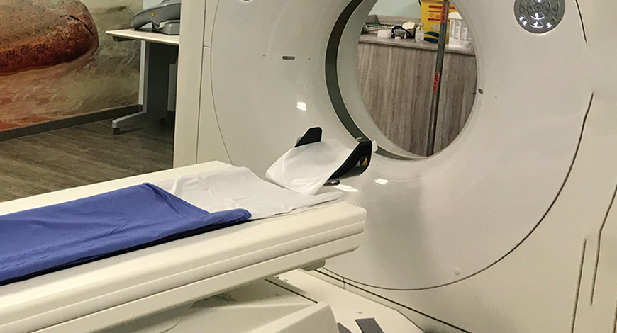
Recent advancements have made CT scanners an essential tool in the diagnostic process, and have paved the way for further innovation in the field of radiology.

Computed tomography (CT) scanning, which started to be developed around 1970, is nowadays the main tool of radiology in daily clinical work. Advances in CT imaging are making scans more accurate, smart, sensitive, and innovative than ever. The CT scan continues to be an invaluable tool in preventive scanning and wellness, especially for the early detection of cancer and heart disease. Advances in medicine and a need for minimally invasive procedures and diagnostics are driving global demand for CT technology to an all-time high. Along with this, recent advances in CT imaging are making them more useful, in more situations.
Greater efficiency in how X-rays are used to create images has resulted in an incredible decrease in radiation dose, along with an increase in detail. New, low-dose machines use tiny beamlets to produce crisp images that can spot abnormalities as small as a grain of rice. These same machines can quickly take detailed images of the beating heart, and assess its health with 3D modeling.
Other advancements are allowing for the isolation of specific elements and compounds, allowing technicians to focus on the most important pieces of the scan. Advances in portable machines and the incorporation of artificial intelligence (AI) have also expanded the usefulness and precision of CT scans tremendously.
Technology trends
Computed tomography scanners are one of the most commonly used diagnostic tools in the field of radiology. Over the years, there have been many advances that have improved the speed, accuracy, and safety of CT scans. Some of the recent technology advancements that are revolutionizing the field of radiology include:
Faster scan times. CT scans have become much faster over the years, reducing the need for patients to hold their breath during scans and increasing the overall efficiency of the diagnostic process. This has been achieved by improving the technology that drives the CT scanner and by using advanced algorithms that reduce the time required to produce images. For example, some CT scanners now have the capability to produce images in just a few seconds, significantly reducing the amount of time required for a patient to complete a scan.
Lower radiation doses. One of the major concerns associated with CT scans is the exposure to ionizing radiation, which has the potential to cause harm to patients, especially children and pregnant women. To address this issue, advances in CT technology have led to a significant reduction in the amount of radiation required to produce an image. For example, some CT scanners are now equipped with technology that adjusts the radiation dose based on the patient’s size and the type of scan being performed. This has made CT scans much safer for patients, especially those who require multiple scans over time.
Improved image quality. Advances in CT technology have also led to improved image quality, making it easier for radiologists to detect small or subtle abnormalities. Improved image quality has been achieved by using more advanced imaging detectors and by using algorithms that enhance the quality of the images produced by the CT scanner. For example, some CT scanners now use algorithms that reduce the amount of noise in the images, making it easier for radiologists to identify small or subtle abnormalities.
Dual-energy CT. Dual-energy CT is a relatively new technology that allows for the simultaneous acquisition of two images at different energy levels, providing more information about tissue composition, and helping to reduce the need for contrast agents. This technology works by using two X-ray sources with different energy levels, which produces two images of the same tissue that can be compared and analyzed. Dual-energy CT is particularly useful for applications, such as evaluating bone density and identifying the presence of certain types of cancers.
Dual-energy CT scanners are a recent development in CT imaging technology that have several benefits over traditional single-energy CT scanners.
These scanners produce images at two different energy levels, which allows for better differentiation between different types of tissue. For example, bones, soft tissue, and metal objects can be easily distinguished from each other in a dual-energy scan, providing improved detail and clarity. Scans can be used to decompose a mixture of materials into its individual components. This allows for the identification of specific materials in a scan, such as metals, plastics, and other foreign objects. Dual-energy CT scans provide a more detailed and accurate picture of the patient’s anatomy, leading to improved diagnosis and treatment. In some cases, dual-energy scans can eliminate the need for additional scans, reducing the amount of radiation exposure and the need for repeat scans. Dual-energy scans can also reduce the number of artifacts in the image, providing a clearer and more accurate picture. Artifacts are areas of the image that appear as distortions or noise and can interfere with an accurate diagnosis. These scans have expanded the diagnostic capabilities of CT imaging. For example, they can be used to diagnose conditions, such as osteoporosis, kidney stones, and certain types of cancer.
Overall, dual-energy scanners represent a major advancement in CT imaging technology, providing improved patient outcomes, and enhanced diagnostic capabilities. This technology is becoming increasingly common in medical facilities, offering patients and healthcare providers a more accurate and detailed view of the body’s anatomy.
Multi-energy CT scanners. Some newer CT scanners are equipped with multi-energy capabilities, allowing them to produce images at different energy levels. This enhances the contrast between different tissues, making it easier to distinguish between healthy and diseased tissues. In recent years, there have been several advancements in multi-energy CT technology, including – multi-energy scanners have become more sensitive, allowing for clearer and more accurate images with lower doses of radiation; advances in image processing algorithms have led to better image quality, reducing noise and improving the resolution of images; spectral CT is another form of multi-energy CT that uses a broader energy range and more advanced detectors to provide a more detailed analysis of tissue composition; and multi-energy CT is being used for a growing range of applications, including cancer detection, cardiovascular imaging, and orthopedic imaging. These advancements have greatly increased the potential of multi-energy CT for improved diagnostic accuracy and patient care.
Several major medical imaging companies have recently launched new models of multi-energy CT scanners, including – GE HealthCare has developed a range of multi-energy CT scanners, including the Revolution CT and the Discovery MI platforms; Philips Healthcare offers the IntelliSpace portal, a multi-energy CT platform designed for use in interventional radiology; Siemens Healthineers has launched a range of multi-energy CT scanners, including the SOMATOM Force platform; Canon Medical Systems offers the Aquilion Prime SP, a spectral CT scanner that uses a range of energy levels to produce detailed images of the body.
These are just a few examples of the many multi-energy CT systems available from leading medical imaging companies.
AI-assisted scans. Another recent development in the field of CT scanner technology is the use of AI software to assist radiologists in identifying abnormalities. AI algorithms are trained on large datasets of images, and can assist radiologists in detecting anomalies that may not be immediately obvious. This has the potential to improve the accuracy and speed of diagnoses, reducing the need for follow-up scans and ultimately leading to better patient outcomes.
CT angiography. The ability to perform CT angiography (CTA) has been added to some CT scanners. CTA is a specialized type of scan that helps in visualizing blood vessels and can be useful for diagnosing conditions, such as aneurysms and blockages. Integration of AI algorithms into CTA systems to automate image analysis has resulted in more accurate results. Due to its non-invasive nature and ability to provide detailed images of blood vessels, CTA is increasingly being used as a diagnostic tool, especially in the fields of cardiovascular and neurovascular disease. The increasing demand for minimally invasive procedures has led to the adoption of CTA as a key tool in interventional radiology and other medical specialties. Hybrid imaging systems that combine CTA with other imaging modalities, such as MRI or PET, have been developed to provide a comprehensive view of the patient’s condition. The adoption of CTA is growing in developing markets due to the improvement of healthcare infrastructure and technological capabilities.
In conclusion, recent advancements in CT scanner technology have significantly improved the speed, accuracy, and safety of CT scans. These advancements have made CT scans an essential tool in the diagnostic process and have paved the way for further innovation in the field of radiology. With continued research and development, it is likely that CT scans will become even more advanced in the future, further improving the diagnostic process, and ultimately leading to better patient outcomes.
Market trends
Artificial intelligence is being increasingly used in CT scanning to improve image quality and reduce radiation exposure. AI algorithms are being used to identify areas of interest in images and automate certain tasks, such as the selection of appropriate scan parameters.
Portable CT scanners are becoming increasingly popular, particularly in emergency and trauma care. These compact, lightweight scanners can be used to quickly image patients in remote locations or those who are unable to move from a hospital bed.
CT scans are being used more frequently to guide interventions, such as biopsies, drainage procedures, and ablations. This has led to the development of specialized CT scanners designed for interventional procedures.
Dual-source CT scanners, which can produce two sets of images simultaneously, are becoming increasingly common. These scanners offer faster scanning times and improved image quality, compared to traditional single-source CT scanners.
CT colonography, a type of CT scan used to detect colon cancer, is becoming increasingly popular due to its accuracy and non-invasive nature. This has led to the development of specialized CT scanners designed specifically for CT colonography.
The demand for CT scanners is growing rapidly in emerging markets, driven by rising incomes, increasing healthcare spending, and a growing awareness of the benefits of medical imaging.
Outlook
Overall, the CT scanner industry is growing rapidly and undergoing significant changes, driven by advances in technology and changing healthcare needs. This is leading to the development of new and improved CT scanners that offer improved patient outcomes and enhanced diagnostic capabilities.


