
Cath Labs
Cardiac cath labs emerging as a game changer
Cutting-edge programs will embrace the catheterization lab’s fresh and revitalized appearance and move on to the next clinically validated option.

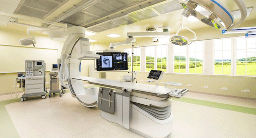
Cardiovascular disease has become a big challenge and has taken the shape of a national epidemic. Earlier, it was presumed to be a disease of the elite class, but lately not so. It is no surprise, therefore, that the need and demand for cardiac services and equipment continues to rise. A cath lab is an integral part of any contemporary large cardiology service. With increasing longevity and percutaneous solutions for several cardiac problems (e.g., TAVR), the utilization of cardiac catheterization services in India is expected to rise even further.
The cath lab industry will flourish with increased number of labs being set up to meet the demand in Tier-II and Tier-III cities. With advent of newer procedures, like mitral clipping and left atrial appendage occlusion, the existing labs will also upgrade and improve imaging quality while minimizing radiation dosages. Labs integrated with IVUS, OCT, and superimposition of CT and TEE images on the cath lab images are starting to gain steam.
Also, newer cath labs are achieving remarkably low radiation exposure without compromising safety. The cath lab equipment industry is poised to grow and is set for exciting times ahead.
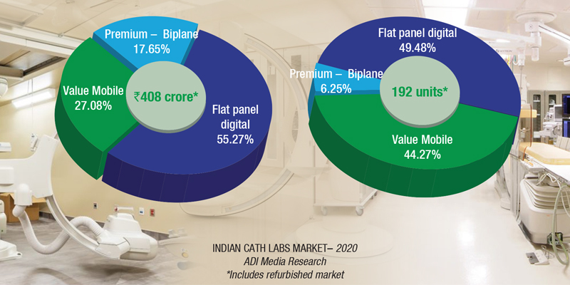
India has about 1600 cath labs in 140 cities. And 225 labs are added every year, albeit 2020 only saw 192 additions. With it being recognized that cardiovascular disease is not only an urban phenomenon, the medical facilities in Indian Tier-III and Tier-IV cities are seeking to set up cath labs, with budgets in the vicinity of ₹6–7 crore. Punjab state government has only two cath labs at the government medical colleges of Amritsar and Faridkot. In the absence of cath labs, cardiac patients in this state have to either pay around ₹150,000 per angioplasty at private hospitals or travel to PGIMER, Chandigarh. And often patients experiencing a heart attack do not manage to reach these facilities so far away from their homes. The government needs to step up here.
World-class cath labs are being set up by private hospitals. In October 2020, Fortis Hospital, Bengaluru, installed one of the most modern biplane cath labs. The cath lab enables the surgeon to see blood vessels of the brain simultaneously in two different planes, the machine is capable of extremely fast 3D acquisition of angiograms and CT scans inside the cath lab, and emergency neuro-interventional cases can now be treated with more precision.
|
Indian cath labs market 2020* |
||
| Tier I | Tier II | Others |
| Philips, GE, and Siemens | Allengers | Shimadzu, Schiller, Zhiem, Skanray, Toshiba, and IITPL |
| *Vendors are placed in different tiers on the basis of their sales contribution to the overall revenues of the Indian Indian cath lab market. | ||
| ADI Media Research | ||
India is the worldwide capital for diabetes and heart disease. It is no surprise that the need and demand for cardiac services and equipment continues to rise. A cardiac catheterization laboratory is an integral part of any contemporary large cardiology service. With increasing longevity and percutaneous solutions for several cardiac problems, the utilization of cardiac catheterization services in India is expected to rise even further.
Recent years have seen a huge array of new procedures performed in the cardiac cath labs and in a variety of similar interventional suites in other areas of the hospital – valve repairs and replacement, closure of a number of congenital heart defects, peripheral vascular interventions for limb and cerebral vascular obstruction, mechanical approaches seeking to prevent arterial embolism from the heart, or dissolving thrombi already present in the peripheral and cerebral arterial circulation.
In the last several years, 3D technologies have advanced tremendously, not only in many medical and surgical disciplines, but also in our daily life outside of work, with an increasing number of new capabilities in smartphones, computer design programs, and movies. 3D pictures are captured in time with 4D imaging.
In cardiology, particularly interventional cardiology, these technologies are highly significant. Understanding the variations in the morphology of veins and the shape of cardiac structures is critical to assuring effective treatments, patient safety, and favorable results.
Newer innovations in both 3D and 4D technologies have recently been created; examples of these developments, as well as how they are emerging as game changers include:
To facilitate structural cardiac surgeries, 3D holograms turn live transesophageal echo imaging into real-time 3D holographic video in the cath lab.
The 3D hologram is shown on a dedicated display screen, and the interventional cardiologist changes the picture orientation, using hand motions and a foot pedal/switch, without disturbing the sterile environment. It also lets the operator to observe the instruments they employ in the cath lab in real time in a 3D format, such as catheters or gadgets. The user does not even need to wear 3D glasses to use this technology.
HeartFlow planner is a virtual tool for coronary artery disease intervention that is noninvasive and real-time. It allows interventional cardiologists to visually map vessels on a 3D coronary tree, with color codes showing fractional flow reserve computed tomography values calculated by a computational fluid dynamics technique for each artery. This appears to be a useful tool for planning percutaneous coronary intervention in arteries with severe disease, since it tries to offer us with a non-invasive method of determining if a stenotic lesion is possibly flow-limiting. CT-FFR, on the other hand, has its own set of limitations, and certain patients may still require invasive FFR for an appropriate evaluation.
For more than a decade, 3D printing has been employed in the surgical profession. It refers to the creation of complicated three-dimensional things using computer-aided design.
In recent years, this technology has become more widely used in structural cardiac surgeries, where 3D models may be produced from a patient’s CT, MRI, or 3D ultrasound pictures. These 3D-printed structures aid procedure planning and device size, as well as allow operators to practice dry runs and pre-procedural navigation.
The modification of these 3D pictures over time is a key component of 4D imaging, which is added to 3D imaging. The direction of blood flow, blood velocities, and shear wall tension are all included in 4D flow pictures. This is also helpful in coronary interventions, structural cardiac surgeries, and other congenital defects, where identifying blood flow in the 4D image is beneficial, especially when the anatomy is complicated. These shifts in location over time assist us in guiding our treatments, not only to assure effective outcomes but also to avoid problems. These 4D pictures need a great amount of data, but they may be generated using either cardiac MRI or computational fluid dynamics, a branch of engineering that combines mathematics and fluid physics. Although 4D imaging is still in its infancy, it is an exciting development in the profession.
These technologies, if developed in partnership with information technology professionals, engineers, and scientists, have the potential to make a difference in a variety of sectors, including coronary interventions and structural cardiac treatments.
Looking back to see how far the industry has progressed is always a worthy endeavor. Indeed, cutting-edge programs will embrace the cath lab’s fresh and revitalized appearance and move on to the next clinically validated option. Issues that need addressing though remain – expensive after-sale-services as well as the need to standardize safety and training practices for cardiac cath lab personnel.


