
CT Scanners
Expanding the boundaries
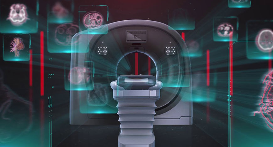
Technological developments in CT have brought significant changes in the speed, spatial resolution, and dose efficiency in newer CT scanners. The advanced and high-end machines have transformed the field of radiology/imaging.

Healthcare is still reeling from the unforeseen disruption that COVID-19 has caused throughout the industry. For some imaging departments, the virus uncovered a disconnect between the imaging technology they have and what they need to thrive. But even without a once-in-a-lifetime pandemic, balancing cost and value has always been a challenge for imaging departments. One reason for this is that technological innovations come at a fast pace, and there is always something new on the horizon that may be a worthwhile investment that expands how you serve patients and referrers – or may be a budget drain with little payoff. Although the fast pace of technological advances is what makes diagnostic imaging such a rewarding field of medicine, it is also what makes it expensive to stay on top of – and indeed, in front of – various trends.
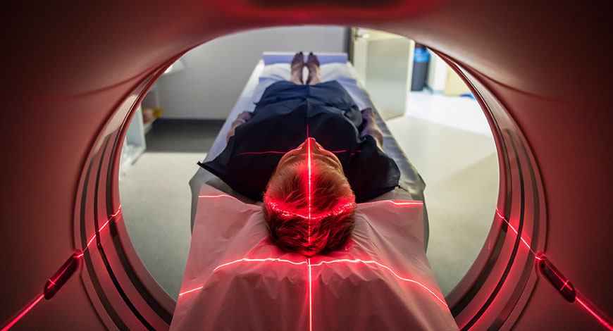
Computed tomography (CT) imaging has broad diagnostic application, and is the imaging gold standard for many clinical indications. However, CT imaging exposes patients to higher doses of radiation than other methods. It carries increased cancer risk for all patients, particularly those in higher-risk categories, such as pediatric, obese, or oncology patients who receive regular screening.
While low-dose and no-dose imaging techniques and modalities exist, a compromise often must be made between patient dose exposure, clinical utility, and cost. Within CT, a direct correlation can be drawn between diagnostic image quality, clinical utility, and radiation dose exposure. Lower-dose procedures produce noisier images, which can impact clinical utility, radiologist productivity, and patient care. Conversely, with increased dose, image quality tends to improve, rendering subtle pathologies more visible – which ultimately benefits radiologists’ diagnostic confidence.
So, how can healthcare providers balance the requirement for high-quality, precise imaging with tight budgets and the need to reduce radiation exposure risk for their patients? New artificial intelligence-based deep learning reconstruction (DLR) and post-processing techniques have recently become available. These methods can consistently improve diagnostic image quality at the lowest attainable dose across all patients and procedures – far beyond what is possible with current reconstruction techniques. This presents a huge potential for imaging organizations to optimize CT imaging programs.
The paradox between improving diagnostic image quality and reducing dose exposure has a complex and generally negative cascading impact on the clinical and operational aspects of CT workflow. Poor-quality images resulting from high noise are more difficult for radiologists to interpret, forcing them to spend more time, carefully reviewing study information. This not only reduces reading efficiency and increases report turnaround time, but it also negatively impacts radiologists’ clinical confidence and the clinical value they can add.
In the late 2000s, iterative reconstruction (IR) was introduced and remains a commonly used technique for improving the quality of CT imaging studies. While IR can reduce image noise and significantly move the needle in terms of dose reduction, IR images can take substantially longer to process. This approach also has limitations in how much dose can be reduced before images take on a blurry or waxy appearance. IR algorithms are also vendor- and scanner-specific. This prevents organizations from upgrading all their CT scanners to IR technologies without sweeping and costly capital equipment replacements. The result has been a phased-in adoption of IR over many years, as CT systems are upgraded or replaced.
Images processed by IR have a different appearance and can introduce distinct artifacts and textures that radiologists have to become familiar with before they can be confident in their clinical diagnoses. While the promise of IR has been largely realized, the limitations of IR cannot be ignored. The industry has now entered the next phase of CT image reconstruction and post-processing, where AI technologies will help to overcome these limitations and push CT imaging into another era.
There are a number of clinical applications that are not well-suited for IR, because of the very low dose and diagnostic requirement for sharp, high-contrast images with fine details.
Older scanners are reliable and well-suited for routine imaging and, therefore, are very common; their inherent limitations make them less suitable for certain patient cohorts. As such, imaging facilities often do not use them for certain advanced procedures or patient types. This can increase patient backlog and wait times, and result in inefficient technologist workload balancing and modality utilization. These older scanners also have fixed costs for electricity, service, and operation, which need to be offset with higher utilization. Any opportunity to increase their clinical utility has important financial and operational implications to the organization.
While some new AI-based CT image reconstruction software is only available on certain premium models of new scanners, other vendor-neutral solutions have emerged in the marketplace. These vendor-neutral solutions are a critical development, because they can be universally applied across all new and older model CT scanners from all vendors. This provides significant financial value by extending the diagnostic life of all CT scanners and deferring capital and professional services costs. It can also improve operational efficiency and reduce patient backlogs and wait times by balancing CT imaging workflow, improving modality utilization and harmonizing image quality across the entire organization.
The application of new AI-based machine learning technologies to CT imaging provides a new variable that clinical and technical stakeholders can use to directly manage both dose and image quality – and, therefore, indirectly all of the subsequent financial, clinical, and operational factors they impact.
AI-based DLR and post-processing techniques are able to process CT images in a matter of seconds – to reduce image noise across a much broader range of doses and exam types than IR.
Image noise, resulting from low-dose CT procedures, causes a negative cascading effect on the quality, efficiency, and cost of imaging services. AI-based DLR and post-processing techniques are able to process CT images in a matter of seconds – to reduce image noise across a much broader range of doses and exam types than IR.
With these techniques, the compromise between dose and quality can be eliminated, delivering clinical, operational, and financial benefits. Because some of these are vendor-neutral technologies, they enable image quality to be harmonized across all CT scanners. Radiologists can more quickly and confidently interpret images without the need to accommodate the image quality variability common today. This holds the opportunity for radiologists to increase their reading productivity, as it could shorten the time needed to read exams coming from certain scanners.
This will likely be more impactful on exams coming from older scanners or challenging exams that are inherently noisier or include more subtle anatomy in pathology. Financially, operational costs associated with inefficient workflow are significantly reduced, capital equipment replacements can be deferred, and reimbursements increase as a result of greater overall productivity.
From the patient perspective, concern regarding the negative effects of radiation can be addressed by leveraging DLR and post-processing to offer screening services that achieve the lowest attainable dose. In doing so, imaging organizations are better positioned to attract new referrals and patients, especially for programs targeted at higher-risk populations, such as low-dose lung screening.
In fact, the recent expansion of low-dose lung screening eligibility creates a significant opportunity for healthcare organizations to take advantage of this new technology.
Early adopters of this new technology have an opportunity to explore the impact on various radiology productivity areas – including (but not limited to) the degree to which the dose can be universally reduced across all CT scanners and patient populations; changes in the conspicuity of subtle pathologies; improved radiologist report turnaround time and clinical confidence; aplicability in ED settings where the processing speed of IR can limit its clinical utility; and financial ROI from increased patient throughput, modality and technologist utilization, and extending the useful life of CT scanners.
The Indian market for CT scanners in 2020 is estimated at ₹1050 crore. The market did not get adversely impacted by COVID-19. After the major dip it saw in April-May-June 2020 quarter, it rapidly picked up in the remaining three quarters of the fiscal. In fact, 2021 is poised for at least ₹1500 crore. April-May-June 2021 have seen a huge uptick in sales, with 60–70 percent being sold in new sites, where CT services did not exist, mainly in Tier-II cities. Big corporate hospitals are also buying more CT machines as a CT scan takes 15 minutes but sanitising the machine after each patient takes 45 minutes.
| Indian CT scanners market – 2020 | ||
| Leading players* | ||
| Tier I | Tier II | Others |
| GE | Siemens | Toshiba, Hitachi, Philips, Medirays, and United Imaging |
| *Vendors are placed in different tiers on the basis of their sales contribution to the overall revenues of the Indian CT scanners market. | ||
| ADI Media Research | ||
However, the preference has shifted toward the value model, with the bulk of demand generating for the 17–63 slice machines, estimated as much as 72 percent of the market. The other segment that has some traction is the 128–160 slices models. The refurbished segment continues to be in demand and contributes about 8 percent to the market by value.
GE continues to dominate the market. This year, apart from Siemens, Toshiba, and Hitachi, two brands that had some visibility were Medirays and United Imaging.
In spite of the Indian doctors cautioning patients not to go in for CT scans indiscriminately, especially in the early stages, hospitals reported patients requesting for a CT scan, where they had classical symptoms of COVID-19 and the RT-PCR test was negative. Also, in cases where a patient in the ICU with severe COVID-19 is not showing any improvement and a chest X-ray shows new lesions, a CT appearance is being done toward a diagnosis of dangerous COVID-19-associated fungal super-infections like aspergillosis or mucormycosis. CT scans also help rule out spontaneous pneumomediastinum and thromboembolism.
The application of new AI-based machine learning technologies to CT imaging provides a new variable that clinical and technical stakeholders can use to directly manage both dose and image quality – and, therefore, indirectly all of the subsequent financial, clinical, and operational factors they impact.
The global CT scanners market had both positive and negative effects of the COVID-19 pandemic. Due to government lockdowns all over the globe, there was no movement for regular check-ups to hospitals. But at the same time, there was an increased inclination toward using a CT scan to diagnose COVID-19. The Centers for Disease Control and Prevention (CDC) recommended against routine CT scans to diagnose COVID-19, based on false-positive rates, scanner contamination, and a lack of change in individual patient management. However, Chinese medical professionals were quick and effective in controlling the disease. Professional medical organizations globally agree that CT plays a crucial role in rapid identification, updated quarantining, and tailoring public health models.
RT-PCR testing’s slow speed and unreliable results became apparent in the early phases of COVID-19 outbreak only. CT scan is far more sensitive and commonly detects the cases of swab misses. However, CT’s higher sensitivity and real-time results come at the cost of increased false positives.
The global CT scanners market is estimated to be worth USD 6.9 billion in 2021. It is expected to reach USD 8.6 billion by the end of 2026, growing at a CAGR of 4.54 percent from 2021 to 2026. 16-slice CT held the largest share of 34.99 percent in the market owing to the fact that 16-slice CT scanners are in high demand for the diagnostic procedures. Moreover, the applications of the 16-slice CT are expanded to wide horizons, thus driving the growth of the market over the next 5 years. 16-slice CT technology has exceptionally advanced and broadened the range of CT applications to include angiography, cardiology, kidney stone evaluation, orthopedics, and more.
A rapid increase in demand for bedside imaging, home healthcare, and increasing CT scan use to evaluate the efficiency of post-interventional medical procedures, medical implants, and anatomical confirmation are key factors propelling the market growth. The rising pervasiveness of diseases, such as cancer, orthopedic, cardiovascular, and dental disorders will also improve the CT scanners market’s growth.
Moreover, a perpetual increase in the number of medical implant operations due to lifestyles and a growing geriatric population base may increase the demand.
Additional drivers of growth include the growing demands for premium healthcare infrastructure from governments. Significant improvements in imaging technologies promise to enhance wellness through rapid and more accurate detection of medical conditions.
A rise in fiscal deficit may hinder the CT scan market growth in developed nations. Moreover, the CT imaging radiation dose continues to be an area of concern. Many patients and medical associations have raised this issue and raised their voices against CT scanning’s possible side effects. The industry focuses on reducing exposure while ensuring the image quality of the CT scan techniques.
Regionally, North America holds the largest market share of around 37.45 percent in 2020 and is expected to hold its position through 2026 in global CT scanners market. This can be attributed to the large number of population suffering from chronic diseases.
Additionally, rapid technological advancements and inclination of people toward newer technologies, coupled with demand for effective treatment procedures, accounts for the largest share of North America.
The global CT market is a consolidated market, owing to the presence of a few major players. The major market players like Philips Healthcare, GE Healthcare, Canon Medical Systems Corporation, and Siemens Healthineers hold a significant share in the industry. Most of the players are focusing on bringing technologically advanced products into the market to acquire the maximum market share.
In July 2021, Canon Medical Systems joined forces with Cleerly in a strategic partnership to support simple and streamlined adoption of cardiac CT. Together, the companies aim to support broader adoption of cardiac CT for the diagnosis and triage of heart disease, including incorporation of AI to improve both the imaging and automate the post processing of datasets.
In May 2021, Philips Healthcare released a workhorse CT system, the Spectral CT 7500, which has regulatory clearance in Europe and from the US FDA. It uses intelligent software to deliver high-quality spectral images on every scan 100 percent of the time without the need for special protocols. The system aims to improve disease characterization, reduce rescans and follow-ups while using the same dose levels as conventional CT scans.
In November 2020, GE Healthcare acquired Prismatic Sensors AB to expand its portfolio of photon-counting computed tomography technology. This technology has the potential to significantly increase clinical performance for oncology, cardiology, neurology, and many other clinical CT applications.
In November 2020, Samsung launched BodyTom Elite and OmniTom, which are mobile whole-body and head CTs, respectively, for point-of-care imaging to determine the ideal treatment strategy for a patient.
The CT imaging radiation dose continues to be an area of concern. Many patients and medical associations have raised this issue and raised their voices against CT scanning’s possible side effects. The industry focuses on reducing exposure while ensuring the image quality of the CT scan techniques.
CT scans help predict COVID outcomes at one month. In a new study published in Radiology of more than 10,000 patients, researchers found that chest CT was 80 percent accurate in diagnosing COVID-19 pneumonia and predictive of death or the need for intubation. Accuracy increased to 86 percent after five days of symptoms. The extent of pneumonia on chest CT was the best predictor of severe outcome (intubation or death) at one month.
Early experience in China and some small studies published at the beginning of the pandemic in early 2020 found CT utility for the modality in confirming COVID infections based on lung scans. Prior to widely available PCR testing for COVID, it was suggested CT could be used to detect the virus, in some cases diagnosing the virus before the patient tested positive using a PCR test. Once the virus reached the United States, there was hesitation to use CT because of its cost, radiation dose, debate over accuracy, and the concerns about the need to decontaminate scanners after each use, which adds to room turnaround times.
However, this new, large-scale, multicenter observational retrospective cohort study confirms the early findings that CT has a high level of accuracy for diagnosing COVID. This study helps answer the conflicting data from smaller, previous studies regarding the diagnostic performance of chest CT for COVID-19 pneumonia.
The STOIC (Study of Thoracic Computed Tomography In COVID-19) project aimed to build a dataset of at least 10,000 CT scans from individuals with suspected COVID-19 pneumonia, evaluated during the first wave of the SARS-CoV-2 pandemic in France.
Using RT-PCR as the reference standard in 10,735 patients with suspected COVID-19 pneumonia, CT was 80 percent accurate and was also predictive of death or need for intubation, the study found.
Researchers looked at data from patients who presented at the emergency departments of 20 French university hospitals between March 1 and April 30, 2020. It included patients who underwent both chest CT and RT-PCR for suspected COVID-19 pneumonia. CT images were analyzed, blinded to initial reports, RT-PCR, demographic characteristics, clinical symptoms, and outcome.
Of the 10,735 patients, 6448 (60 percent) had a positive RT-PCR result. With RT-PCR as reference, the sensitivity and specificity were 80.2 percent and 79.7 percent, respectively, with strong agreement between junior and senior radiologists. Of all the variables analyzed, the extent of pneumonia on CT was the best predictor of severe outcome at one month. A score, based solely on clinical variables, predicted a severe outcome when it also included the extent of pneumonia and coronary calcium score on CT.
Readers classified CT scans as positive or negative for COVID-19, based on criteria published by the French Society of Radiology. Multivariable logistic regression was used to develop a model predicting severe outcome (intubation or death) at one-month follow-up in subjects positive for both RT-PCR and CT, using clinical and radiological features. If the first RT-PCR result was negative, but turned out to be positive in the following seven days after CT, the patient was considered as positive for SARS-CoV-2 infection. CT studies in China often found evidence of COVID pneumonia in the lungs from early infection prior to positive PCR tests, and this was also found in the study.
CT has undergone substantial advances in technology in the past decades; it has significantly improved cancer diagnosis, reduced the length of hospitalization, and helped preparation for surgeries. The advances in the CT scan technology in recent years include reducing harmful radiation to patients, enabling faster scan speed, as well as improved image quality.
Scanning radiation exposure. Lowering radiation emission has long been a challenge for the CT scanning scientists, as radiation doses for CT scans are significantly higher than conventional X-rays and can cause serious long-term health risks to patients. These days, there are novel scanning techniques that dramatically reduce the radiation during scanning. According to a source, University College of London, date July 23, 2020, title New CT scan method lowers radiation exposure from Science News, a CT scan technique that splits a full X-ray beam into thin beamlets can deliver the same quality of image at a much-reduced radiation dose, according to a new UCL study. The article adds that the use of beamlets enables a sharper image resolution, as the part of the scanner reading section, is able to locate where the information is coming from more precisely. This CT scan technology can deliver the same quality of image at a much-reduced radiation dose.
In addition, the new CT detector technology, which measures the amount of radiation transmitted through the body, provides more accurate level of radiation exposure.
Higher-slice CT systems. When the 64-slice CT scanners were introduced in the early 2000s; they were quickly purchased to replace the older 16-slice CT scanners, and became the new standard for CT technology. The current 32- or 64-slice scanners are up to four times faster than the original generations of scanners. OHSU Knight Cardiovascular Institute offers a 256-slice CT scan diagnostic imaging equipment, which takes super-fast pictures, in milliseconds, of a moving heart using a three-dimensional tool format on a computer monitor. It also provides much more visual details about the heart’s function and structures. The increased speed of scanning enables physicians to provide more accurate diagnosis and better treatment for early or advanced coronary artery disease.
Spectral CT imaging. Spectral CT technology (also called dual-source/dual-energy CT) is another trend that is becoming more and more integrated into major CT vendors’ technologies. Spectral CT breaks down X-ray photos by chemical elements, based on viewing one part of the body at two different kV energies, with a dual-source CT scanner. Instead of scanning a patient several times using different energies to focus on different tissue types, spectral CT technology provides different views from a single scan. This software can also highlight and remove chemical compounds solely based on their atomic number, i.e., iodine, calcium, and metals. This can create contrast and non-contrast images created from one single scan. This technology is particularly useful in the treatment of gout and locating kidney stones; it also improves metal artifact reduction. The spectral CT imaging and detector systems have led to a reduction in dose by 70–82 percent over the last few years.
Artificial intelligence. The field of artificial intelligence (AI) is transforming medical imaging. In computed tomography (CT), AI holds the promise of enabling further reductions in patient radiation dose through automation and optimization of data acquisition processes. Optimization of image-reconstruction parameters, and image-denoising methods improve image quality. New AI-based deep learning reconstruction (DLR) and post-processing techniques improve diagnostic image quality.
Computed tomography texture analysis (CTTA) obtains biomarkers that provide quantitative assessment of tumor heterogeneity by analyzing the differences and patterns within the pixel values of an image. CTs can be worked with as a matrix of numbers, which can reveal patterns and characteristics. There are over twenty parameters, which are standardized in radiomics.
Mobile and portable CT scanning. Portable CT scanning is clinically and economically beneficial. Physicians can limit the risks associated with intra-hospital transport, such as the compromise of monitoring equipment, intravenous lines, or intubation tubes. Portable CT scanners offer fast CT deployment to hospitals and radiology units and have been especially beneficial to treatment of head injuries, by reducing the risk to patients during transportation and increased risk during the treatment process. In addition, minimizing the need for extra transport contributes to economic benefits, as well as improves the use of other equipment and enhances the overall quality of patient care.
When it comes to efficiently mobilizing CT for COVID-19 imaging, a highly effective strategy can be literally mobilizing scanners to enable movement throughout the hospital. By adopting mobile CT equipment, facilities can keep infected patients apart from the general imaging population through a policy of CT distancing, so to speak.
Transporting infected patients to radiology departments can disperse germs throughout the hospital and even turn the radiology waiting room – which accommodates patients from across the hospital and beyond – into an extremely dangerous place. In particular, distancing is important for vulnerable intensive care patients, who are at higher risk for infection and often deadly disease progression.
Mobile scanners also provide a simple, fast, and relatively cost-effective way to expand CT capacity for the increased demands of infectious disease imaging without the complex and costly demands of siting and construction of additional imaging suites. In addition, they can be more easily adapted to meet other imaging needs when the immediate need for infectious disease imaging decreases – and it will.
Besides distancing the infected and non-infected patients, mobile scanning can help solve significant related challenges of altered workflow and scanner disinfection. The recommended necessary disinfection of scanners and the imaging suite following a CT exam of a suspected COVID-19 patient is an arduous process – far more extensive and time-consuming than for general imaging procedures. If cleansing is not thorough, patients may become infected. This process significantly changes department routines and hampers patient throughput, which in turn affects exam scheduling for all patients, along with department revenues. Maintaining a coronavirus-dedicated CT will allow time for scrupulous cleaning without impacting routine department workflow on other devices.
Going mobile can not only help facilities in the fight against COVID-19 today, it can also broaden capabilities and move radiology deftly into the future.
An important new trend is photon-counting CT (PCCT) technology, which has the potential to define a new standard of performance for premium CT systems for many years to come. PCCT has the promise to further expand the clinical capabilities of traditional CT, including the visualization of minute details of organ structures, improved tissue characterization, more accurate material-density measurement or quantification, and lower radiation dose.
Several vendors are working on photon-counting detectors for their next-generation CT scanners. PCDs also allow the binning of photons of different energies, so all CT images created can be post-processed into high- or low-energy images, or spectral imaging. Some vendors say this detector technology will increase data-mining capabilities from multiple-energy detection, and contribute to the improved utility and efficacy of AI applications.
While this technology has been researched academically for several years, it appears several vendors are close to commercializing these detectors. This technology will be a hot topic in 2021 and beyond.


