
Industry
Helium-The liquid gold of MRI
Geopolitical factors, shipping and supply chain issues all affected the availability of helium over the summer of 2022. The situation has somewhat eased now, albeit prices for the coolant have skyrocketed.

About a quarter of the helium consumed worldwide is used for the extreme cooling of superconducting magnets, and demand is on the increase. At the same time, helium is a rare element – at least here on our planet. Produced by the decay of various radioactive elements in the Earth’s crust, it either accumulates in rocks or percolates into pockets of natural gas, of which it is a common constituent. From there, helium is brought to the surface by volcanic activity or must be recovered from natural gas using complex separation techniques.
Largely a non-renewable and non-recoverable natural resource, if it’s vented into the atmosphere, it is so light that it escapes the earth’s gravity and boils off into outer space. The problem is that conventional MRI scanners are capable of venting a lot of it. Helium is used in an MRI scanner to cool the machine’s magnet so that the electrical current in its coil can flow indefinitely without encountering resistance – a state known as ‘superconductivity’. A conventional MRI scanner can boil away as much as 1,500 liters depending on its fill level, and once it’s gone, it’s gone for good. With aging populations and the steady growth of neurological and oncological disease worldwide, the number of global MRI scans also continues to grow at a significant rate thanks to their superiority in contrast resolution and tissue differentiation over any other diagnostic imaging modality. As a result, more and more helium is required to meet these demands, making the practice effective, but not environmentally sustainable. Coupled with geopolitical tensions affecting helium supply chains, there is currently a world shortage of the precious gas and prices are rising. In early 2022, maintenance shutdowns at gas production centers in the U.S. and Qatar and fires at a new Russian facility disrupted the supply of helium further.
The manufacturers have recognized this profligate use of helium in MRI scanners is unsustainable. Several vendors have introduced so-called “helium-free” MR systems. is in short supply, so it is expensive.
These helium-free systems use very low amounts of helium, the helium system is completely sealed and no vent or quench stack is required. This significantly reduce costs of stack installation and allows MRI rooms to be located anywhere, without the need to consider how to vent the system outside the building.
Permanent magnet MRIs from 0.2T to 0.4T do not require any liquid helium for cooling. Higher field MRI systems ranging from 0.5T to 1.2T require less liquid helium than 1.5T MRI scanners. Some systems are great at maintaining the low temperature, allowing very little or no helium to boil off. Hence their name, zero boil-off MRIs. They are also called 4K systems as they maintain the temperature of 4 Kelvin, which ensures that helium remains in its liquid state, rather than turning into gas and dissipating.
While there are a few commercialized helium-free systems on the market, they cost more than traditional systems, which limits their full potential for adoption. This is especially true in developing countries, where upfront costs are a primary driver for adoption.
Also, manufacturers have been putting helium reclamation units on all MRI scanners since the late 1990s.
Indian market dynamics
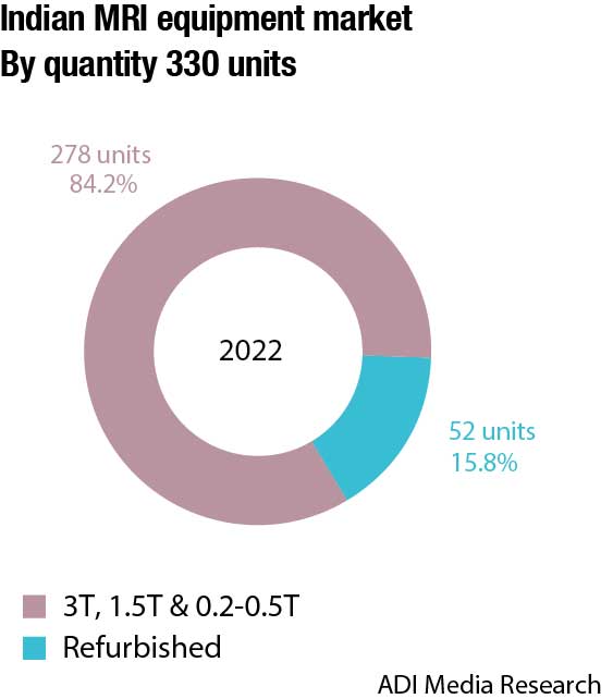
The Indian MRI market in 2022 is estimated at ₹1515 crore, and 330 units. This is a 15 percent decline over 2021 by value and a 13.7 percent increase by number of units. This can be explained by the increase in sales to tier 3 and 4 cities, that had never procured MRI machines. As Covid receded, and CT scanners lost their feverish relevance, the smaller hospitals took stock, and saw they had made major financial gains in the two-year period. In an attempt to differentiate their facilities and to equip with the latest machines, briskness in demand was observed for MRI machines, albeit a larger percentage was for pre-owned. And this is where brands as Phantom, Medirays, and Sanrad stepped in. These vendors imported the three leading brands Philips, Siemens and GE pre-owned machines. Those who did not buy the pre-owned machines opted for the 0.2T- 0.5T segment, that saw a jump in market share from 0.55 percent in 2021 to 5.3 percent in 2022 by value.
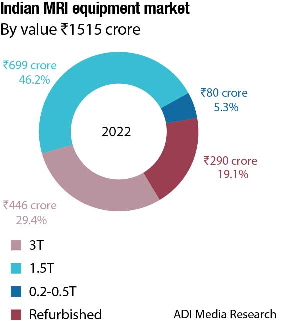
Subsequently 3T machines declined from a 41 percent share in 2021 to a 29 percent share in 2022. 1.5T continued to command a 47 percent market share by value. This is in stark contrast to the 2021 market trends when the two premium segments, 3T and 1.5T systems, dominated the market with a combined 88 percent share by value, and 80 percent share by numbers, and only seven units in the 0.2T–0.5T segment were bought in 2021.
Having said that, now that the machines in metros are more than ten years old, from 2023 onwards, it is expected that there will be a demand for 3T machines from the hospitals here.
| Major players*– 2022 | |||
| Tier 1 | Tier 2 | Tier 3 | Others |
| Siemens, Philips, and GE | Phantom, Medirays, and Sanrad | United Imaging | Fuji (Hitachi) and Canon (formerly Toshiba) |
| *Vendors are placed in different tiers on the basis of their sales contribution to the overall revenues of the Indian MRI market. | |||
|
ADI Media Research |
|||
Global market
The global MRI equipment market is estimated to reach USD 8.36 billion by 2027, from USD 6.52 billion in 2022, growing at a CAGR of 5.1 percent.
Low field strength MRI machines have field strength lower than 1.5T, mid-field MRI machine have field strength ranging from 1.5T up to 3T, and high field MRI machine has a field strength of more than 3T. The midfield strength segment, machines ranging from 1.5T up to 3T held the largest share in 2022 and the trend is expected to continue. They provide a good balance between capturing images with higher precision and affordable pricing.
The high-field strength segment, more than 3T, is expected to experience the fastest growth rate. Various research studies are being conducted to understand the efficiency of high-field strength MRI machines in various clinical applications. Some players have already started developing high-field strength MRI machines, and are receiving approval from the regulatory body. Currently, 7T MRI machines are being studied only for brain and knee imaging due to the lack of advanced coils required for other clinical applications. The development of advanced coils to widen the application of 7T MRI machines is expected to propel growth.
Key drivers. Academic institutes, research laboratories, hospitals, and physicians are all interested in advances in MRI techniques, such as superconducting magnets, open architecture, high-field MRI, and software applications. Furthermore, introducing MRI-compatible pacemakers will considerably expand the number of eligible MRI treatments. The increasing demand for rapid and effective diagnostic treatments will likely drive MRI machine usage. Various paramagnetic contrast agents, such as gadolinium-DTPA, are injected intravenously to provide sharper, more precise, and accurate images in less time. In coming years, the MRI market is predicted to grow further with the increasing development of intraoperative MRI and its use in different applications, such as neurosurgery.
Advancements in diagnostic techniques including open MRI, visualization software, and superconducting magnets are fueling the growth of the MRI market. The recent advances seen in MRI technology are mainly on the software. Moreover, the development of cardiac pacemaker-compatible MRI systems is also expected to propel the market growth in the cardiology segment. Aged people usually suffer from various problems, such as cardiovascular and neurological disorders, which fuels the use of MRI and promotes market growth.
Diffusion and diffusion-tensor imaging with tractography, neuroimaging comprising MR spectroscopy, perfusion imaging, and functional imaging utilizing the bold approach are likely to drive market growth throughout 2023–2027. Because high-magnitude MRI systems are becoming more popular, different colleges are doing research or studies on them. This machine is intended to pave the way for new ways to diagnose diseases, including Alzheimer’s, heart disease, diabetes, and cancer. Technological advancements in MRI systems, such as 7T, a high-resolution MRI, will improve the comfort of imaging the interior structure and functioning of the CNS while reducing processing time. Because of this, players in the MRI systems market will see considerable growth opportunities. Through new product releases, partnerships, and training experts for various MRI systems, healthcare businesses primarily focus on delivering enhanced healthcare facilities and goods to boost their economic opportunities.
Challenges. The market’s growth is projected to be limited by the high cost of MRI systems, the incompatibility of MRI systems in some patients, and declining reimbursement rates for MRI therapies. In addition, the growth is hampered by delayed product approval, a lack of skilled professionals, and frequent product recalls owing to the industry’s strict regulatory structure. However, the global MRI market is expected to be showcasing a healthy growth rate in the coming years due to the growing adoption of new normalization and decreasing impact of Covid-19.
Region insights. The MRI market in North America accounted for the largest share in 2022, followed by Asia-Pacific. The rising prevalence of chronic diseases in this area, such as breast cancer, cardiovascular disease, and neurological disease, drives up demand for imaging analysis. Because of the United States’ well-established healthcare system, and the activities being carried out by numerous organizations, this area is anticipated to expand its market share in the future. In North America, Siemens and Philips are the leading brands.
In the Asia-Pacific region, the market is estimated to be the fastest-growing region owing to the growing improvements in the healthcare infrastructure and increasing healthcare spending. With a fast-developing healthcare service business and many qualified healthcare workers, India, China, and Japan provide growth opportunities.
The European MRI market is predicted to hold a considerable share of the global market. The growing disposable income from UK, Germany, Italy, and France, increasing developments in the healthcare infrastructure, and growing demand for imaging technologies, are propelling the market.
Recent advancements and future of MRI

Dr TBS BUXI
Chairman, MRI & CT Scan Department,
Sir Ganga Ram Hospital
Magnetic resonance imaging (MRI) has remained at the pinnacle of diagnostic imaging since its innovation. MRI will continue to evolve by generating superior quality of patient images by incorporating artificial intelligence, sustainable hardware, and patient-centric workflows. Emerging technologies and innovations in MR are aimed at better healthcare outcomes at lower cost of care, with improved patient and clinician experience.
In my opinion, MR innovations will continue to focus on helium-free MRI magnets as the sustainable, small-footprint, and lightweight solution to provide improved diagnostic outcomes. This is also an urgent need of the hour to address the ongoing severe shortage of helium that would probably get worse with time in next few years, and will create serious problem for conventional super-con MRI system users in the long run. Currently, helium-free 70-cm bore size super-conducting MRI magnets are available up to 1.5T – we can expect that even the higher-field MRIs would also become helium-free in near future! Those environment friendly sustainable MRI systems of future will incorporate increased field of view, digital coils, increased gradient strengths for advanced studies, AI-augmented hardware and software features optimized for patient throughput, comfort, and superior clinical outcome.
As the latest trend, we have observed that MR-compatible monitoring and biopsy devices are becoming commercially available that are enabling new diagnostic pathways by providing comprehensive and precise diagnosis. For an example, recently developed Doppler ultrasound-based MR gating device to facilitate gated fetal cardiac MR studies (like Smart-Sync in Philips MRI) can enable robust diagnosis of fetal congenital heart diseases during fetal MR scan. Commercially available MR-guided breast and prostate biopsy solutions, along with UroNav like commercially available system for ultrasound-MR fusion biopsy, are also becoming more user friendly for radiologists to adopt these advanced methods in their routine practices.
Further advances in AI-powered software engines for image acquisition and reconstruction will keep delivering superior-quality MRI images in less time, making MR scanning even faster and more efficient. Future MRI machines will be equipped with intelligent systems, providing high-speed, high-resolution images delivering confident and precision diagnoses even in complex diseases. AI will continue to play a major role in improving overall scanner workflow from MR image acquisition and reconstruction, post processing, and generation of reports. AI-powered workflows will integrate intelligence, guidance, and ease-of-use, resulting in enhanced scheduling efficiency and faster scanning, with improved patient and clinical staff comfort and thereby improving productivity of MR department.
In summary, we can expect sustainable and AI-powered MRI machines of near future will provide patient-centric scanning with very high quality of clinical MR images, AI-assisted decision support for enhanced diagnostic confidence, and improved clinical outcomes that will benefit both patients and radiologists.
Market players. A few of the notable companies operating are Hitachi Medical Corporation, Toshiba Medical Systems, Siemens Healthcare, GE HealthCare, Imris Inc., Fonar Corporation, Esaote S.P.A., Neusoft Medical System, Aurora Medical Imaging, and Philips Healthcare.
What’s new in MRI technology
Recent advances in MRI machines promise to overcome the limitations of traditional scanners, allowing doctors to produce clear images of more body parts in more people.
Increasing the scan speeds. Nicholas Dwork, PhD, assistant professor in the Department of Biomedical Informatics at the University of Colorado, School of Medicine, has filed a provisional patent for a technology that could increase scan speeds of three-dimensional magnetic resonance imaging (MRI). The invention could lead to faster results, increase the clinical applications of MRIs, and ultimately improve patient care.
Complex mathematics and engineering are involved in generating images of internal organs and tissues when patients enter the magnetic tube of an MRI machine. Any movement by the patient can corrupt the images, and the scan can take an hour or longer. During this time, the machine makes loud noises as radio waves reverberate off bodily structures to create images. Because the field of view – the part of space that is imaged – is typically rectangular, the radio waves also bounce off areas outside the body. This is where Dwork’s technology comes into play. His method adjusts the sampling pattern created by the machine’s magnetic fields.
New sensor uses MRI to detect light deep in the brain. Using a specialized MRI sensor, MIT researchers have shown that they can detect light deep within tissues, such as the brain.
Imaging light in deep tissues is extremely difficult because as light travels into tissue, much of it is either absorbed or scattered. The MIT team overcame that obstacle by designing a sensor that converts light into a magnetic signal that can be detected by MRI. This type of sensor could be used to map light emitted by optical fibers implanted in the brain, such as the fibers used to stimulate neurons during optogenetic experiments. The fiber they used is similar to those used for optogenetic stimulation, so this kind of sensing could be useful to researchers who perform optogenetic experiments in the brain.
With further development, it could also prove useful for monitoring patients who receive light-based therapies for cancer, patients receiving treatments that involve light, such as photodynamic therapy, which uses light from a laser or LED to kill cancer cells.
The researchers are now working on similar probes that could be used to detect light emitted by luciferases, a family of glowing proteins that are often used in biological experiments. These proteins can be used to reveal whether a particular gene is activated or not, but currently they can only be imaged in superficial tissue or cells grown in a lab dish.
Noninvasively tracking propagation of brain signals on millisecond timescales. A team of researchers affiliated with multiple institutions in the Republic of Korea has developed a new way to use MRI for noninvasive tracking of the propagation of brain signals on millisecond timescales. In their paper published in Science, the group describes how they developed the new technology, its features, and how well it worked when tested on mice.
MRI works by sending a magnetic field along with radio waves through tissue to produce images of the tissue under study. It works because different types of tissues have different properties. MRI is also used in different ways, depending on the part of the body under study. Blood-oxygen-level-dependent functional MRI is used to obtain images of the brain of a living person – the technique allows for observing changes in blood flow in the brain that serve as a stand-in for an increase in neuronal activity. In practice, blood-oxygen-level-dependent functional MRI produces multiple images over time, generally over several seconds. In this new effort, the researchers have modified the way an MRI brain scan is performed.
The new technique, called direct imaging of neuronal activity (DIANA) works by modifying a traditional MRI machine to create images faster – on the millisecond scale – potentially at the speed of thought. It does so by creating partial images that are combined afterward to create a high-resolution image that represents an instance of brain activity. After a single scan, multiple high-resolution images are created that show the state of the brain as it responds to a stimulus, such as a jolt of electricity applied to the whiskers of test mice. By generating images so quickly, the technique allows researchers to observe the propagation of brain signals from one part of the brain to another in a noninvasive manner.
The new technique has thus far only been tested in mice, but the researchers are already calling it a game-changer, suggesting it could transform the way that scientists study the brain – and could lead to a new understanding of how it works.
Outlook
As medical technology advances, MRI technology is continually improving to provide more accurate, faster, and safer scans, ultimately benefiting both patients and medical professionals. Also, advancements in MRI technology have reduced the likelihood of adverse reactions to the contrast agents used in some MRI scans.
Overall, these advancements in MRI technology have led to better patient care. Medical professionals are now able to make more informed decisions, based on the data collected from MRI scans. This technology has also helped to reduce the need for more invasive procedures and surgeries, as the non-invasive nature of MRI scans can often provide enough information to diagnose and treat patients.
As the field of medical technology continues to evolve, it is expected that further advancements in MRI technology will be made in the future.


