
X-ray Equipment
Imagining more possibilities with DR
Dynamic digital radiography is the next evolution in x-ray. This is a technology worth exploring.

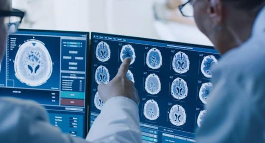
Digital radiology may represent the greatest technological advancement in medical imaging in the last decade. The use of radiographic films in x-ray imaging might become obsolete in a few years. An appropriate analogy that is easy to understand is the replacement of typical film cameras with digital cameras. Images can be immediately acquired, deleted, modified, and subsequently sent to a network of computers.
The benefits of digital radiology are enormous. It can make a radiological facility or department filmless, eliminating chemical processing of films. The referring physician can view the requested image on a desktop or a personal computer and often report in just a few minutes after the examination was performed. The images are no longer held in a single location, but can be seen simultaneously by physicians who are kilometers apart. In addition, the patient can have the x-ray images on a compact disk to take to another physician or hospital.
The advantages of digital x-ray systems have played a key part in their adoption; their speed and accuracy, as well as quick processing times, allow for significantly higher patient screening volumes than earlier. This has pushed companies to focus on product development and innovation. However, these systems are priced at a premium, which slows their greater adoption. Other factors, such as declining reimbursements, lack of infrastructure, particularly in developing and underdeveloped countries, and potential risks, associated with radiation exposure, are also expected to hinder the growth of this market.
Global market
The global digital radiography (DR) market size was estimated at USD 4.5 billion in 2022, and is expected to hit around USD 9.4 billion by 2030, with a notable CAGR of 9.66 percent during 2022 to 2030.
The portable system segment dominated the DR market in 2022. Factors like the growing quantity of diagnostic imaging due to the aging population and the widespread deployment of portable radiology systems throughout diagnostic centers. In addition, an increase in ailments, such as vascular, dental, and cancer, particularly breast disorders and others, is propelling the portable DR segment forward. The government’s support for numerous radiology research programs is also propelling the sector forward.
The stationary system segment is the fastest-growing segment of the market in 2022. The rise in the prevalence of chronic diseases and cancer is driving the expansion of the stationary digital radiography system market. Many patients, particularly in developing nations, have imaging tests every year. Furthermore, future population expansion will increase demand for digital radiological detectors, boosting the segment’s growth. One of the significant digital radiography market trends is that older persons acquire age-related diseases, necessitating the usage of a variety of radiographic tests.
Technology insights. The direct digital radiography (DDR) segment dominated the market in 2022. DDR is a type of x-ray examination that produces a digital radiologic image on a supercomputer very quickly. This technique captures information during object examination, using x-ray subtle plates, which are then transferred to a processor without the obligation of the middle cassette. A detector sensor alters the incident x-ray radiation to an equal electric control, which is then translated to a digital image.
The computed radiography (CR) segment is the fastest-growing segment in 2022. When imaging plates are subjected to x-rays or gamma rays, the energy of the incoming radiation is recorded in a specific phosphor layer in computed digital radiography. The latent picture from the plate is then read out using a scanner, which stimulates it with a very precisely focused laser beam. The plate produces blue light with an intensity proportionate to the quantity of radiation acknowledged during the contact when triggered.
A major benefit of DR-based systems over film and CR systems is that images are sent directly to a computer screen from the x-ray imaging plate, thereby eliminating the need to retrieve the image plate for processing images or taking a second x-ray shot. An effective dynamic range, better medical evaluation, superior and accurate image capture, reduced radiation exposure for patients and workers, flexibility in image management, improved patient throughput, superior evaluation of data and images, and lower operational costs are some of the other advantages of DR systems. Due to these advantages, the use of DR systems is on the rise.
North America dominated the market and accounted for the largest revenue share of 30 percent in 2022. The market is being influenced by factors, such as increasing adoption of modern technology and improved healthcare infrastructure, strong purchasing power, and a fair reimbursement framework.
In the Asia-Pacific, the market for DR systems is estimated to witness the fastest CAGR during 2023 to 2030 owing to the increased demand for better imaging devices and supportive government initiatives for improving healthcare infrastructure in the region. Rapidly developing economies and improving healthcare services in Southeast Asian countries, China, Japan, and India are expected to propel growth. For instance, according to Enhance International’s Sam Radwan, China’s estimated healthcare spending in 2050 might be greater than Germany’s overall GDP in 2020. Furthermore, the increasing prevalence of chronic disorders and the rise in the geriatric population is expected to boost the market growth in Asia-Pacific.
The key players are developing high-end mobile DR systems, which gained popularity since the Covid-19 pandemic, as well as integrating Al into these imaging systems, which have demonstrated impressive accuracy and sensitivity in the detection of imaging abnormalities. As a result, it is being more widely adopted and used in the field of medical imaging.
Some of the prominent players in the x-ray systems market include Fujifilm, Carestream, Mindray Medical, Philips, GE Healthcare, Siemens Healthineers, Canon Medical Systems, Shimadzu Corporation, Hologic, Inc., New Medical Imaging, and Agfa.
What is new in digital radiography?
A standard x-ray provides tremendous value to a clinician for its ability to quickly provide a static image of anatomical structures. There are limitations, however, to static exams. Multiple conventional x-rays are often needed to show a joint in flexed and extended positions. A single static image cannot provide the details of how a joint moves between flexion and extension points or tell the complete story.
Dynamic digital radiography (DDR) adds dynamic, or motion, capability to x-ray technology that captures sequential radiographic images in a single exam, enabling clinicians to observe the dynamic interaction of anatomical structures in motion over time. Unlike fluoroscopy, x-ray with DDR, is not viewed in real time; so an exam can be performed by a radiologic technologist without a physician present in the exam room.
DDR is an enhanced version of a standard digital radiography system that acquires up to 15 frames per second for as long as 20 seconds, resulting in a maximum of 300 x-ray images with a dose equivalent to about two standard x-rays. With DDR, radiation is lower than fluoroscopy or CT, and requires a shorter exam time than MRI.
Applications in pulmonology. The benefits of DDR are being explored across a variety of disciplines. In pulmonology, DDR can be used to visualize and quantify lung function in relationship to surrounding structures. Radiologists can watch the patient’s muscles as they breathe – as they inhale and as they exhale. DDR opens up potential for artificial intelligence and various analytic applications to process the exam, quantify movement, and track changes over time. It is taking the simple chest x-ray and pairing it with high-end technology to now produce a functional exam that was not present previously.
Applications in orthopedics. In orthopedics, DDR can be used to assess instability, musculoskeletal injury, sources of pain, and treatment follow-up of any joint throughout its range of motion – whether it be the neck, spine, shoulders, or knees. It can also be a helpful tool for postoperative evaluation of movement in place of a more expensive CT or MRI exam.
Other applications. DDR use is also being explored as an effective tool for other applications, including swallow studies for speech therapy, or as an alternative to fluoroscopy for assessing diaphragmatic movement during sniff tests. Clinicians can now see more with a dynamic x-ray. Because DDR is another x-ray technique using the same x-ray system, the equipment and technician workflow is the same. It just adds an extra minute of time. DDR is available on the same digital radiography system that performs conventional x-ray exams, providing a cost-effective solution for hospitals and practices.
For many physicians, DDR provides a more complete diagnosis and has become a key differentiator for their practice. It introduces a technology with clinical value that also has the benefit of a CPT code for reimbursement.
In the hospital environment, DDR adds value to what can be done. For patients, there is a wow factor when they see the motion in their x-ray. Anecdotal feedback suggests that patients who see a dynamic x-ray are more likely to understand their condition and adhere to their rehabilitation plan.
Adding movement gives new insight, which makes it easier for clinicians to adopt technology and make it useful. There is clinical value, economic value, technological value and patient appeal. This is a technology worth exploring.
Industry updates
In December 2022, Carestream Health announced new innovations in its long-length imaging digital radiography (DR) detector that are aimed at improving patient comfort, productivity, and efficiency. The Carestream DRX-LC Detector (Pending FDA 510(k) clearance) captures long-bone and spine images with a single exposure, making it ideal for pediatric imaging and long-bone and spine images of people with limited mobility. In contrast, the traditional multi-shot approach requires three to five exposures followed by manipulation and stitching of the images. There are several key advancements in this next-generation long-length detector. The first is compatibility with DR mobile systems and mobile retrofits, in addition to x-ray rooms and room retrofits. The mobility of the system and its X-factor capability allow facilities to use the same detector across multiple DRX systems for all phases of surgical treatment, from pre-planning imaging in the radiology department to capturing images during the procedure in the OR to confirm placement, and for post-operative assessment. The versatile detector can be wireless or tethered. The wireless option eliminates trip hazards in the OR and minimizes the risk of contamination. The DRX-LC detector has a Cesium (CsI) scintillator for better dose efficiency.
In November 2022, with an eye on helping radiologists to become more efficient, collaborate, and avoid burnout, Philips focused on its new artificial intelligence (AI) and imaging informatics offerings at RSNA 2022. In digital x-ray, Philips shined the spotlight on its Fluoroscopy 7000 N – ProxiDiagnost N90 digital radiography/fluoroscopy unit. The 2-in-1 system can be utilized in applications, such as chest, full leg and spine, upper and lower extremities, and skull exams. In addition, it is suitable for gastrointestinal studies, arthrography, venography, lymphography, myelography, and digital subtraction angiography.
In November 2022, plans for a new US subsidiary and a range of new product launches were among the highlights for Canon Medical Systems at RSNA 2022. The company unveiled a number of new DR offerings in its booth, including its CXDI-Pro and CXDI-Elite series of wireless detectors and built-in automatic exposure control (AEC) assistance technology. In addition, Canon introduced its Intelligent NR technology for use on the CXDI series of control software. The company noted that the CXDI-Elite series will be the first of its digital x-ray imaging systems to utilize built-in AEC Assistance technology, which automatically ends exposures without the use of an additional receptor. AEC Assistance works via both wired and wireless communication.
In November 2022, display firm LG Business Solutions USA showcased its two prototype display monitors and a prototype digital x-ray detector at RSNA 2022. One of the new monitors is a 55-inch 4K in-plane switching (IPS) surgical monitor with 650 units of brightness. Its largest surgical monitor designed to date features multiple inputs and outputs to accommodate flexible installation scenarios, antireflection and antifingerprint coatings, a 178-degree viewing angle, and protection against liquid and solid foreign objects.
In September 2022, Konica Minolta Healthcare Americas received US Food and Drug Administration clearance for its Chiropractic Straight Arm (CSA) x-ray system, featuring dynamic digital radiography (DDR). CSA is a compact system and features an array of designs to optimize workflow, increase efficiency, and improve outcomes. DDR was developed by Konica Minolta to enable the visualization of anatomy in motion so that clinicians can interpret the dynamic interaction of anatomical structures, such as tissue and bone, with physiological changes over time. The incorporation of DDR in the CSA system will further enhance the diagnosis and management of musculoskeletal conditions.
In August 2022, ViviX-S F series of digital radiography (DR) detectors by Vieworks was cleared by the US Food and Drug Administration. The ViviX-S F series includes cassette-sized static x-ray DR flat-panel digital detectors that come in three sizes, including 25×30 cm (ViviX-S 2530FW), 36×43 cm (ViviX-S 3643FW), and 43×43 cm (ViviX-S 4343FW). Technical specifications of the detectors include a 99µm pixel size; a glass-free, thin-film transistor design; and a semidynamic multiframe mode. It also includes image-processing technology with the company’s VXvue software-based scatter correction.
In July 2022, Fujifilm Healthcare Americas launched FDR Cross, a new portable fluoroscopy and DR system for hospitals and ambulatory surgery centers. FDR Cross features a battery-powered dual-function C-arm and offers portable fluoroscopy and radiography on a single platform. The technology was designed to reduce the need to bring in additional equipment for image-guided procedures. FDR Cross includes a pivoting tube head and removable detector, compatibility with Fujifilm’s FDR D-EVO III detectors in multiple configurations, an imaging area of up to twice the size of standard C-arm systems when using the 17×17-inch detector, and an interchangeable detector holder, among other features.
Radiographers split on perceptions of AI in practice
Radiographers are split on whether they feel prepared to start using new artificial intelligence (AI) technologies in daily practice, according to a survey published December 18, 2022, in the Journal of Medical Imaging and Radiation Sciences.
A team led by Dr Theophilus Akudjedu from Bournemouth University in England found that the jury is out on how confident radiographers are in using AI, but most agree that teaching AI technologies should be included in the radiography curriculum.
“There are many benefits of AI-enabled radiography workflows and improvement on efficiencies, but equally there will be widespread disruption of traditional roles and patient-centered care, which can be managed by a well-educated and well-informed workforce,” Akudjedu and colleagues wrote.
Clinical practices have progressively implemented AI, including in the field of medical imaging. Radiologists and radiographers have experienced a surge in such applications, with AI helping reduce clinical workloads as an alternative reader. But the investigators pointed out that not everyone has the education and training needed to fully understand AI’s benefits and risks.
Akudjedu and colleagues sought to assess the knowledge, perceptions, and expectations of radiographers working across various settings on the use of AI technologies. To do so, the group conducted a survey that included 314 radiographers – most reported practicing in North America.
Out of the total survey participants, 170 reported not using AI or related technologies daily, and 107 stated they had six to 10 years of radiography practice experience.
The survey found the following:
- 67.6 percent of respondents either agreed or strongly agreed that AI will change daily clinical radiography practice.
- 32.8 percent reported that they expected AI to reduce the workload of the radiography workforce.
- 50.3 percent of respondents reported feeling well-prepared to implement AI into daily practice.
- 31.4 percent reporting being somewhat confident in using AI technologies or innovations in their daily practice.
- 47.3 percent either disagreed or strongly disagreed that there are enough AI training opportunities currently available for radiographers.
- 69.3 percent either agreed or strongly agreed that learning about AI technologies should be included in the radiography teaching curriculum.
The survey indicates an overall lack of understanding when it comes to AI-specific language in radiology, according to the researchers.
“[Our work] shows that, while a considerable proportion of the worldwide radiography community expressed willingness to accept and adopt AI in practice, they, however, lack the theoretical knowledge or experience to do so,” they concluded.
Way forward
Addressing the common challenges that conventional x-ray brings, DR is paving the way for high-quality, safe, and reliable imaging in 2023 and beyond.
Additionally, 2023 appears to be a watershed year for medical image management. Radiologists will experience less stress and burnout as a result of moving to the cloud, relying more on teleradiology, and seeing a rise in AI and machine learning, which will also assist to improve work-life balance. The patient experience will also be improved by new technologies, providing improved access to care for those who are underserved and accelerating diagnosis and treatment.


