Nuclear Medicine Equipment
India poised to produce medical isotopes

Upcoming research and development efforts will lead to an even wider array of indications for nuclear medicine both in diagnosis and treatment.
The field of nuclear medicine has been involved in a significant technological advancement, and in a tremendous progress in radiochemistry, that opened up new and fascinating diagnostic and therapeutic opportunities. The leap forward of stand-alone systems beside hybrid imaging design improvement and novel theragnostic methods development has made possible a wider use of nuclear medicine techniques in several diagnostic scenarios.
With technological advancements in the field of imaging, there has been a significant evolution of hybrid systems in the past decade. The major advancement in this regard is that of combined PET modalities. The innovation of PET/CT modality has been so successful that the major imaging system manufacturers do not provide standalone PET systems any longer. Based on the success of the hybrid PET/CT systems, hybrid PET/MRI and SPECT/CT systems have also gained attention. These hybrid systems can provide precise images with better resolution compared to standalone systems. They can provide morphological as well as physiological information in just one examination. For instance, in the case of skeletal evaluation, a SPECT/CT system offers accurate localization, along with improving the specificity of the information provided by CT. Due to advantages, such as these, many hospitals are now replacing their standalone systems with hybrid systems.
The COVID-19 outbreak has significantly impacted the availability of hospital resources worldwide. This has been primarily managed by dramatically reducing in-patient and outpatient services for other diseases and implementing infection prevention and control measures. Both diagnostic and therapeutic nuclear medicine procedures declined precipitously, with countries worldwide being affected by the pandemic.
Most of the nuclear medicine scans and therapies are carried out in outpatient departments as follow-up cases, whereas new studies are usually dealt with in inpatient departments after going through COVID-19 screening. Nuclear medicine lacks portable SPECT and PET scanners, and there is a constant need to inject the patient with radiopharmaceuticals so the movement of the patient is not restricted. Hence it is very crucial for nuclear medical workers to have a thorough knowledge of precautionary measures to prevent COVID-19 transmission. A majority of heart patients who go through nuclear cardiology procedures are usually above 60 years of age and carry other extreme health risks, such as diabetes, hypertension, and chronic renal and lung diseases.
The Indian SPECT and PET industry use isotopes in scans. Tc-99m is critical for SPECT scans because it emits photons in the form of gamma rays, while other positron-emitting isotopes are used for PET scans.
At present, India produces all major isotopes in the country under the aegis of BARC and also imports some from Europe, Australia and other Asian countries, it said, adding the planned PPP can transform India’s stature in the global nuclear medicine industry.
Bhabha Atomic Research Centre (BARC) has announced that it has “evolved” the design for the country’s first public-private partnership research reactor for production of nuclear medicines. The state-run the Department of Atomic Energy (DAE) will share the technology of production of a variety of nuclear medicines and the private entities will get exclusive rights to process and market the isotopes produced in the research reactor, in lieu of investing in the reactor and processing facilities. The proposed reactor is expected to come online within five years of the beginning of the construction. The construction is planned to start after obtaining all requisite permissions.
The large scale and the technology being deployed for the planned research reactor will enable India to not only become a significant global player in the growing nuclear medicine market. This project will be a major step towards making India self-reliant in key radio isotopes used in medical and industrial applications. As a result, it will increase availability of effective and affordable treatments for cancer.
Seventeen companies, including those from India, have shown preliminary interest in partnering with DAE for setting up the research reactor. The DAE will be bearing the cost of constructing the reactor upfront and expects the partner to get a high return on capital invested. Preparatory work, including design and regulatory clearances is moving at good pace and a formal Request For Proposal (RFP) for selection of the private partner will be initiated in the December quarter.
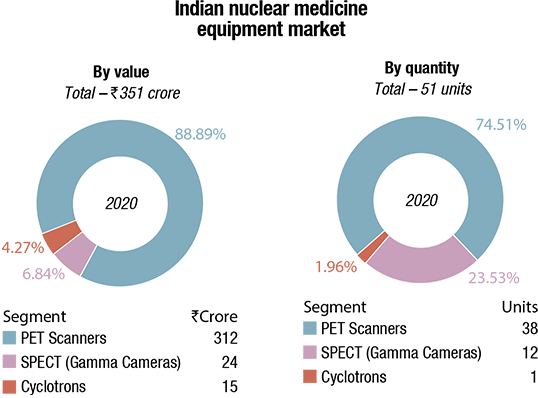
The Indian market for nuclear medicine equipment in 2020 is estimated at ₹330 crore. The PET-CT scanners continue to dominate the segment, with a 74.5 percent share by volume, and a 88.8 percent share, by value.

Thirty-eight PET-CT scanners were procured in 2020, with the government buying only five units this year. These were purchased by the Tamil Nadu government, under a private partnership agreement issued through five companies. One high-end digital scanner was purchased by Deenanath Mangeshkar Hospital and Research Center, Pune; it was from Siemens. The remaining machines were at an average unit price of ₹8 crore each, all bought by private facilities. The buyers included two units to Anderson Diagnostics & Labs, one unit to Manipal HealthMap Diagnostics at Kasturba Hospital, Manipal, and one unit to Matrix Diagnostics Center, Bengaluru.
The gamma cameras with SPECT tomography and integrated CT scanners segment have a 23.52-percent share by volume, and a 6.84-percent share, by value, at 12 units, estimated at ₹24 crore.
One cyclotron was sold by GE, at about ₹15 crore to GetWell Poly Clinic & Hospital, Jaipur. A multi-specialty diagnostic center, the facility has a cutting-edge technology nuclear medicine department. Ranjan Kabra, taking his father’s legacy of quality without compromise forward, was responsible for acquiring the first 64-slice PET-CT in the state of Rajasthan, when he established this department.
In India, GE dominates the segment and shares the space with Siemens. The refurbished machines also have a miniscule share. While 2019 saw the entry of PET-CT scanners in Tier-II and Tier-III cities, with basic models, in 2020 the buying pattern shifted to high-end machines. Almost a similar number were procured; however, the value was about 20 percent higher.
Gamma cameras saw a decline in numbers, while the average unit price remained the same. This was largely because the government buying was just one unit in this period – the funds were targeted at devices directly used for treatment of COVID-19.
The demand has picked up since July 2021, and the vendors have had a good 3Q and 4QFY22.
Amid the COVID-19 crisis, the global market for nuclear medicine imaging equipment estimated at USD 2.3 billion in the year 2020, is projected to reach a revised size of USD 2.9 billion by 2027, growing at a CAGR of 2.9 percent, estimates Research and Markets. Hybrid positron emission tomography (PET) systems are projected to record a 3.5 percent CAGR and reach USD 1.3 billion by 2027. The single-photon emission computed tomography (SPECT) systems segment is expected to grow at a CAGR of 2.7 percent for the next seven-year period.
The nuclear medicine imaging equipment market in the US is estimated at USD 629.6 million in the year 2020. China, the world’s second largest economy, is forecast to reach a projected market size of USD 585.4 million by the year 2027, trailing a CAGR of 5.5 percent. Among the other noteworthy geographic markets are Japan and Canada, each forecast to grow at 0.7 percent and 2.2 percent respectively over the next seven years. Within Europe, Germany is forecast to grow at approximately 1.4 percent CAGR.
In the global planar scintigraphy segment, USA, Canada, Japan, China, and Europe will drive the 1.8 percent CAGR estimated for this segment. These regional markets, accounting for a combined market size of USD 434.6 million in the year 2020, will reach a projected size of USD 494 million by the end of 2007. China will remain among the fastest growing in this cluster of regional markets. Led by countries, such as Australia, India, and South Korea, the market in Asia-Pacific is forecast to reach USD 393.6 million by the year 2027, while Latin America will expand at a 3 percent CARG.
Nuclear imaging systems are priced at a premium and require high investments for installations, which increases the procedural cost for patients as well. This affects the adoption rate of new systems, especially in emerging countries; most healthcare facilities in these countries, consequently, cannot afford such systems. In emerging countries like Brazil, the average cost of a PET system is between USD 400,000 and USD 600,000 while a SPECT system would cost between USD 250,000 and USD 400,000. These high costs, coupled with maintenance costs, are expected to hamper the market growth. In addition, shorter half-life of radiopharmaceutical and shortage of radionuclide Tc99m (technetium 99) also create challenges for the market growth.
Healthcare facilities that purchase such costly systems often depend on third-party payers (such as Medicare, Medicaid, or private health insurance plans) to get reimbursements for the costs incurred in the diagnostic, screening, and therapeutic procedures performed using these systems. As a result, factors, such as continuous cuts in reimbursements for diagnostic imaging scans and the increasing cost of nuclear imaging systems are preventing medium-sized and small healthcare facilities from investing in technologically advanced nuclear imaging modalities.
Data-integrated imaging systems enable the processing and reconstruction of images, computer-assisted recognition of medical conditions, generation of 3D images, and the use of appropriate quality-control systems. With the help of data integration, physicians can easily compare the scans to effectively observe the progression of the disease. To effectively devise a treatment plan, clinicians are demanding access to integrated, comprehensive data on the patient’s diagnostic history. In addition to data integration, there lies a huge opportunity on making data available through mobile technologies. This will help doctors to view and study scans wherever they are. Due to the advantages and convenience of integrated systems and a huge demand from clinicians, companies are focusing on developing systems with integrated technologies.
Prominent players operating in the global nuclear imaging equipment market are Siemens Healthineers, Koninklijke Philips, GE Healthcare, Toshiba Medical Systems, Neusoft Medical Systems, Mediso Medical Imaging Systems, CMR Naviscan Corporation, Digirad Corporation, SurgiEye GmbH, and Positron Corporation.
These key players are implementing multiple strategies to maintain their significant share in the market. The strategies implemented include product developments, business expansion, and collaborative development.
In November 2020, CMR Naviscan Corporation acquired certain Gamma Medica (GM) assets and intellectual property. This acquisition positions CMR Naviscan to provide a comprehensive solution for the diagnosis and care of breast-cancer patients.
Opportunity for emerging industry participants. As per the World Health Organization’s (WHO) definition, nuclear medicines are the specialized medicines containing radioisotopes, intended to be used for diagnostics and therapeutic purposes. High activity and long-term adverse effects increase their demand, which imposes stringent production and distribution regulations on these medicines.
The increasing R&D activities in this industry are fostering influx of new molecules as well as the new treatment options for various conditions, such as respiratory disorders, thyroid disorders, and bone diseases. These emerging applications of nuclear pharmaceuticals are anticipated to open avenues for market players, hence ensuring future growth potential.
Over a period of time, about 100 radioactive molecules have been discovered and their diagnostic as well as therapeutic applications unleashed. In the last two decades, the number of PET radiopharmaceuticals has increased almost exponentially. At present, it is possible to label the pharmaceutical compound with either a γ or positron-emitting radionuclide for diagnostic applications (SPECT or PET), or a ß-minus, or α radionuclide emitter for therapeutic purposes. This concept is the fundamental basis of theranostics. The 21st century has been seeing the development of new radiopharmaceuticals for theranostics to reshape personalized medicine with expanding indications, mainly in neuroendocrine tumor and prostate cancer patients.
Though this industry is evolving and there is swift growth in the market revenues, the players are facing several challenges related to production.
Some of these challenges include shorter shelf-life of nuclear medicine, which restricts its use and distribution; the production process unlike traditional pharmaceuticals is complex and manufacturers have to bear additional cost to maintain the nuclear reactor; and furthermore, currently the production is at a smaller capacity, compared to conventional medicines, complying with cGMP, QA and QC standards.
Considering these challenges and presence of few players operating at global nuclear medicine market level, molecules, such as Technetium-99m, which is used most widely, are facing severe shortage.
So far, the focus has been on diagnostic aspects of nuclear medicine; however, this focus is shifting toward therapeutic areas of these molecules. Emerging applications in the field of oncology, thyroid, bone metastasis, and lymphoma are expected to open up opportunities in the aforementioned treatment areas. For instance, advent of radio-labeled antibodies enabled targeted therapy in treatment of cancer. Radio-labeled antibodies only attach to target site and provide radiation effect directly to the cancer cell.
Though this industry has many entry barriers, rising interest and awareness is anticipated to attract many players in the nuclear medicine market with heavy investment in developing newer solutions. During the last decade, the market saw the emergence of new radionuclides, and the number of products under development has grown considerably. An increasing interest from private investors and conventional pharmaceutical industries for the nuclear medicine market was observed, of course, mainly in the therapeutic area. This led also to some major M&A activities. During the past six years, over USD 16 billion were invested in M&A transactions in the radiopharmaceutical industry. Pharmaceutical companies are investing in radiopharmaceuticals with therapeutic purposes, which may reach estimated revenue of USD 13 billion in 2025. New opportunities for development, investments, mergers, and acquisitions are just popping up.
The major players in radiopharmaceutical market include Eckert & Ziegler Group, Mallinckrodt Pharmaceuticals, GE Healthcare, Bracco Imaging S.p.A, Nordion, Inc., NTP Radioisotopes SOC Ltd., and Eczacibasi-Monrol.
 Kalpesh Bhatt
Head of Molecular Imaging,
Siemens Healthineers, India
Proper management of cancer treatment requires accurate diagnosis and evaluation of the spread of the disease. Imaging technology plays a crucial role in addressing the need for precise and early treatment in oncology. PET/CT scanner provides sophisticated imaging to assess the original tumor as well as the metastatic disease.
PET/CT technology has evolved over the years and improved its performance with the advanced technology. Time-of-flight (ToF) technology has been established as a gold standard since the last few years, and with innovation in hardware, performance of ToF has improved, providing clinical images with higher resolution, detectability at lower radiation dose, and faster scanning. With increasing cancer load in India, demand for ToF PET/CT has increased even in remote cities in India, improving the management of cancer. patients.
Siemens Healthineers offers unique technologies like FlowMotion and Multiparametric Imaging, enabling tailored protocols basis clinical requirement of patients.
PET versus SPECT. PET and SPECT are the leading nuclear medicine modalities. These techniques have their roots not too far into the past. SPECT has emerged as a highly effective technology for tracing numerous funcis due to two main aspects, by means of even more advantageous features of the device in terms of temporal and spatial resolution and the high effectiveness of 18F-fluorodeoxyglucose (FDG) as marker of metabolic activity of cancerous tissue in the era of cancer research. One would in fact wonder if it is PET that decreed the success of the 18F-FDG or the opposite. It has long tional aspects of the body, such as vascular flow, metabolism, and neural transmission. PET technology has undergone a more rapid and intense development since the 1980s. This acceleration been discussed, and still is debated, which technique between PET and SPECT is superior in research and in the clinical arena.
Indeed, both methods will be necessary in the future due to different strengths and limitations. One of the strengths of SPECT is the availability and the lower costs. PET is superior due to better resolution, more sophisticated tracers, not only FDG and because it is quantitative, and absolute quantification with SPECT is a limitation as compared to PET. However, it is very likely that both techniques will still be used for different indications.
The strong interplay between research on disease-development mechanisms and on organ-damage identification is the key element of the recipe for success of these nuclear medicine tools. Several applications in oncology, cardiology, and neurology are expressed by the use of PET and SPECT imaging. In addition, different physio-pathological patterns of systemic diseases can be investigated by both methods. In particular, the theragnostic approach to oncological patients finds in the use of novel tracers and advanced scanners its natural aptitude and its reason to be. Instead, cardiology has the great merit to have driven and directed the development of solid-state gamma cameras that are now the big asset of all nuclear medicine fields. With regard to neurology, a key role for the use of PET and SPECT scanners derives from the huge effort in radiopharmaceutical research with introduction of several imaging probes tracing specific receptor activities at the service of the more fascinating and unexplored medicine field.
The success of these instruments is amply demonstrated by the literature and by the significant diagnostic improvement that the use of these devices has made possible both in terms of accuracy and time to reach diagnosis. This potential is further highlighted by the continuous push of research on industry for technological implementation. In particular, we have seen the realization of two strands. On the one hand, it has introduced the conception of dedicated gamma cameras that would allow faster acquisition times with a lower dose of administered radiation and greater comfort for the patient. These advances have materialized in hardware lighter in design and in size, accommodating large amount of data acquired in list-mode. On the other hand, we have assisted in the integration of stand-alone nuclear medicine methods with morphological techniques in hybrid devices. The resourcefulness of nuclear medicine instruments is documented not only from a more strictly technical point of view with reference to the devices used but also with respect to the radiopharmaceuticals used.
In fact, the improvement of the acquisition capacity related to different photon-emission spectra has made possible the simultaneous acquisition of data concerning the decay of different radionuclides at the same time, paving the way for the development of acquisition techniques with dual-tracer administration to investigate different pathophysiological patterns in a single acquisition. This opportunity together with the continuous introduction of new molecules with specific diagnostic targets for the development of molecular probes that are increasingly personalized on the patient, provided several applications in oncology, neurology, and cardiology as well as in the study of systemic disease with multi-organ involvement at different levels. On the other hand, in addition to the growth of the nuclear medicine tools potential, interdisciplinary knowledge at the service of imaging has further amplified the diagnostic and prognostic rule of the acquired data.
The artificial intelligence approach is one of the big challenges of future imaging. Next-generation experts will have to deal with this powerful application. In other words, the market is approaching an era where every single acquired pixel can be the key to read the past, the present, and the future of each disease, and even when the instrumens is not available to understand data, the very near future may offer novel opportunities to give meaning to information obtained in the past. In this context, PET, SPECT and hybrid modalities may greatly benefit interdisciplinary knowledge to improve their diagnostic, prognostic, and theragnostic potential but, at the same time, need to be ready to face the new-era challenges, providing faster, more accurate, and non-invasive methods to get as many row data as possible in a very short time at the lowest possible radiation exposure.
Strengths and limitations of these two modalities are continuously changing in the context of ongoing hardware and software development, and they need to be constantly understood and updated. It becomes clear that the potential overcoming of the historical limitations of each technique needs to be highlighted with appropriate resonance. At the same time, any improvement in terms of image reconstruction algorithms has to be made available for the scientific community so that all researchers may apply innovative tools to state-of-the art scanners.
The industry has arrived at a crossroads in nuclear medicine. Its success and future depends on further innovation of radiopharmaceuticals, hardware equipment, regulatory acceptance, and approval.
The development and success of new radiopharmaceuticals is expected. Onsite labelling, using generators, will positively affect clinical practice within nuclear medicine departments.
Both PET and SPECT technologies are rapidly evolving, embedded in hybrid devices, and the adjunction of MRI makes the field even more versatile. Multimodality imaging will continue to enhance PET use in the coming years. The evolution of PET systems will further improve significantly in terms of sensitivity and spatial/timing resolution with the continuous improvement of scintillators and electronics. Also, total-body scanners, dual-probes, and optical imaging may bring significant benefits to diagnostic accuracy, radiation exposure, and cost-efficiency, ultimately leading to highly personalized imaging and subsequent treatment.
Finally, theranostics encompasses two worlds, introducing the concept of personalized and targeted-treatment into state-of-the-art clinical routine. Further accelerated developments and wider use of radiolabeled nuclide therapies is likely to happen in the years to come within the 21st century.


