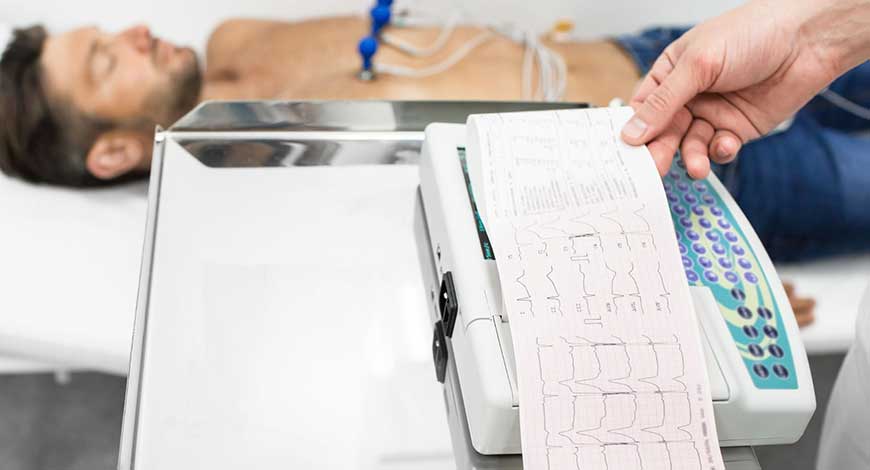
ECG Equipment
Integrating AI into ECG

The implementation of the AI-ECG is still in its infancy, but a continuously growing clinical investigation agenda will determine the added value of these AI tools, their optimal deployment in the clinical arena, and their multifaceted and so-far largely unpredictable implications.
The electrocardiogram (ECG) is a ubiquitous tool in clinical medicine that has been used by cardiologists and non-cardiologists for decades. Although the acquisition of ECG recording is well standardized and reproducible, the reproducibility of human interpretation of ECG varies greatly according to levels of experience and expertise. In this setting, computer-generated interpretations have been used for several years. However, these interpretations are based on predefined rules and manual pattern or feature recognition algorithms that do not always capture the complexity and nuances of an ECG. However, artificial intelligence (AI) in the form of deep-learning convolutional neural networks (CNNs), which have been used mostly in computer vision, image processing, and speech recognition, has now been adapted to analyze the routine 12-lead ECG. This development has resulted in fully automated AI models mimicking human-like interpretation of ECG, with potentially greater diagnostic fidelity and workflow efficiency than traditional rule-based computer interpretations.
Indeed, ECG is an ideal substrate for deep-learning AI applications. The ECG is widely available and yields reproducible raw data that are easy to store and transfer in a digital format. In addition to fully automated interpretation of ECG, rigorous research programs using large databanks of ECG and clinical datasets, coupled with powerful computational capabilities, have been able to demonstrate the utility of AI-enhanced ECG as a tool for the detection of ECG signatures and patterns that are unrecognizable by the human eye. These patterns can signify cardiac disease, such as left ventricular (LV) systolic dysfunction, silent atrial fibrillation (AF), and hypertrophic cardiomyopathy (HCM), but might also reflect systemic physiology, such as a person’s age and sex or their serum potassium levels. Among several potential clinical applications, a single 12-lead ECG might, therefore, allow the rapid phenotyping of an individual’s cardiovascular health and help to guide targeted diagnostic testing in an efficient and potentially cost-effective manner.
One of the top priorities for the application of AI to the interpretation of ECG is the creation of comprehensive, human-like interpretation capability. Since the advent of digital ECG more than 60 years ago, ongoing effort has been toward rapid, high-quality, and comprehensive computer-generated interpretation of ECG. The problem seems tractable; after all, ECG interpretation is a fairly circumscribed application of pattern recognition to a finite dataset. Early programs for interpretation of digital ECGs could easily recognize fiducial points, make discrete measurements, and define common quantifiable abnormalities. Modern technologies have moved beyond these rule-based approaches to recognize patterns in massive quantities of labelled ECG data.
Several groups have worked to create AI-driven algorithms, and some of these algorithms are already in limited clinical use. Some studies have developed CNNs from large datasets of single-lead ECGs and then applied them to the 12-lead ECG. For instance, using 2 million labelled single-lead ECG traces collected in the Clinical Outcomes in Digital Electrocardiology study, one group used a CNN to identify six types of abnormalities on the 12-lead ECG. This study demonstrated the feasibility of this approach, but widespread implementation or external validation in other 12-lead ECG datasets is forthcoming. Another group conducted a similar study of the application of CNNs to single-lead ECGs and demonstrated that the CNN could outperform practicing cardiologists for some diagnoses. However, whether this approach will translate to clinically useful software for 12-lead ECG interpretation remains to be seen.
Nevertheless, although great progress has been made toward a comprehensive, human-like ECG-interpretation package, the realization remains on the horizon. Even in its most modern incarnation, the package lacks the accuracy needed for implementation without human oversight. Additionally, computer-derived ECG interpretation has the potential to influence human over-readers and, if inaccurate, can serve as a source of bias or systematic error. This concern is particularly relevant if the algorithms are derived in populations that are distinct from those in which the algorithms are applied. This limitation underscores the need for a diverse derivation sample (ideally including a diverse patient population and varied means of data collection that reflect real-world practices), rigorous external validation studies, and phased implementation with the ongoing assessment of model performance and effectiveness. Quality control systems, based on regular over-reads by expert interpreters of ECGs, who are blinded to the output of the CNN, will allow continuous calibration of the model.
AI algorithms can be applied to wearable technologies, enabling rapid, point-of-care diagnoses for patients and consumers. Although many algorithms have been derived using 12-lead ECG data, some studies have demonstrated favorable performance even when algorithms are deployed on single-lead ECGs. The performance of AI-ECG algorithms for the detection of HCM or the determination of serum potassium levels, when applied to single-lead ECGs, has been shown not to be significantly different from the performance when applied to 12-lead ECGs. Additionally, signals other than the ECG can also be analyzed using AI approaches. For instance, a deep neural network has been developed to detect AF passively from photoplethysmography signals obtained from the Apple Watch. Newer iterations of the Apple Watch now allow users to confirm the presence of AF using electrophysiological signals obtained through a single bipolar vector.
The Covid-19 pandemic has highlighted the need for rapid, point-of-care diagnostic testing. For instance, early interest in the use of hydroxychloroquine or azithromycin for the treatment of the infection resulted in a marked increase in the use of these medications. Given the potential for these medications to prolong myocardial repolarization and increase the risk of dangerous ventricular arrhythmias, the FDA issued an emergency-use authorization for the use of mobile devices to record and monitor the QT interval in patients taking these medications. The FDA also issued an emergency-use authorization for the detection of low LVEF as a potential complication of Covid-19, using the AI-ECG algorithm integrated in the digital Eko stethoscope. Furthermore, an international consortium is currently evaluating the ECG as a potential means to diagnose Covid-19, cardiac involvement or the risk of cardiac deterioration, given the known ECG changes and cardiac involvement in patients with Covid-19. Although results are not yet available, these types of investigations emphasize the potential power of digitally delivered AI technologies for timely deployment at the point-of-care and large-scale implementation.
In contrast to the data obtained through the clinical history, medical record review, or imaging tests, the ease and consistency with which ECG data can be obtained and analyzed for the development and implementation of AI models is likely to accelerate the uptake of AI-ECG in clinical applications, with ensuing increases in workflow efficiency. The demonstrated capabilities of the AI-ECG have the potential to influence the spectrum of patient care, including screening, diagnosis, prognostication, and personalized treatment selection, and monitoring. The preliminary data on the performance of AI-ECG algorithms are clearly promising, but these technologies will be meaningful only in as much as they improve our clinical practice and patient outcomes. To this end, several AI technologies are currently being tested in various clinical applications.
The AI-ECG technologies offer great promise, but it is important to acknowledge several potential challenges. Given that models are often derived from high-quality databases with meticulously obtained ECGs and well-phenotyped patients, their application to ECGs obtained in routine clinical practice in real-world settings might be poor. Similarly, although the models might perform well in one population, they require rigorous evaluation for external validity in diverse populations.
Clinical and technical challenges also exist to the application of AI-ECG to patient care. AI analysis relies on standardized digital ECG acquisition that is not universal in current clinical environments, even in high-resource settings. Poor-quality data can prevent the effective development and application of AI-ECG technologies. Current AI tools cannot analyze ECGs stored as images, which limits generalizability in some clinical environments. Furthermore, the infrastructure to integrate AI-ECG results into electronic health records and make them available at the point of clinical care is not widely available.
 Dr Amit Bhushan Sharma
Associate Director and Unit Head Cardiology,
Paras Hospitals
SCD is responsible for half of all heart-related deaths. Role of ECG cannot be over emphasized either in diagnosing ailments with increased risk of SCD or predicting future events so as to take preemptive measures. Most common causes of SCD include CAD (ST-T changes), HOCM (LVH with secondary ST-T changes), DCMP (nonspecific ST changes), long QT (more than 480 ms) and short QT (less than 320 ms) syndromes, WPW syndrome (short PR and delta wave), hyperkalemia (tall T wave, long PR interval, wide QRS and sine wave), hypokalemia (VT), hyper, and hypomagnesemia. Certain rare causes include channelopathies, such as Brugada syndrome (ST elevation in right precordial leads) and CPVT (polymorphic VT). ECG abnormalities are obvious predictor of SCD in future. Common abnormalities that predict future SCD are QT internal, early repolarization, QRS duration, LVH, and T wave inversion, QRS-T angle, and heart rate variability. Severe AS or PS patients may be at high risk of SCD. ECG will show LVH/RVH with secondary ST-T changes. Congenital septal defects with eisenmenger physiology are at increased risk of SCD with ECG of RVH. Certain diseases may cause SCD after bradyrrhythmia, such as IWMI, hyperkalemia, degeneration of conduction system, toxic substances, and myocarditis (ST-T changes). ECG as the most basic tool, when used judiciously, timely, and interpreted correctly, goes a long way in helping clinicians suspecting such patients very early and averting the undesired events of future.
The true potential of deep-learning AI applied to the ubiquitous 12-lead ECG is starting to be realized. The utility of the AI-ECG is being demonstrated as a tool for comprehensive human-like interpretation of ECG, but also as a powerful tool for phenotyping of cardiac health and disease that can be applied at the point of care. The implementation of the AI-ECG is still in its infancy, but a continuously growing clinical investigation agenda will determine the added value of these AI tools, their optimal deployment in the clinical arena, and their multifaceted and so-far largely unpredictable implications. As with any medical tool, the AI-ECG must be vetted, validated, and verified, and clinicians must be trained to use it properly, but when integrated into medical practice, the AI-ECG holds the promise to transform clinical care.


