
Urinalysis Instruments and Reagents
New technology meets clinical knowledge
As analytical variation has been greatly reduced, more efforts need to be focused on the pre-analytical phase.

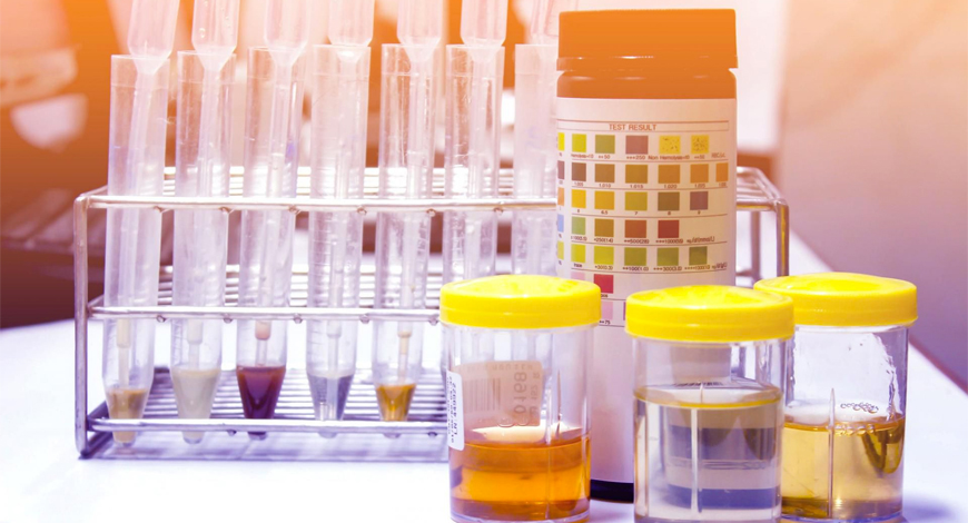
Urinalysis has been a useful diagnostic tool since thousands of years. Although urine was the first body fluid to be examined by mankind for the diagnosis of diseases, it is still one of the most common specimens used in clinical and diagnostic laboratories. Urinalysis includes visual examination, evaluation of chemical parameters, and microscopic examination. Although manual microscopic techniques are standardized, the traditional microscopic examination of the urinary sediment is labor intensive, time consuming, imprecise, inaccurate, and has wide interobserver variability. Therefore, automatic urine analyzers for high-volume laboratory settings were developed in order to improve the certainty of quality measurements, and save labor and time.
Modern technologies and informatics have all but eliminated many manual aspects of urinalysis, which now work in concert with advanced methodologies, such as flow cytometry in automated microscopy and urine dilution for improved interpretations. Additional methods, such as MALDI-TOF mass spectrometry for rapid pathogen identification, are now part of the urinalysis analyzer’s growing arsenal.
Several manufacturers have developed automated urine analyzers, which integrate and perform all urinalysis components for use in clinical laboratories. There are two main types of analyzers, based on different technologies. The first type is an image-based microscopic urine sediment analysis that uses a video camera to capture and sort particles according to preset particle dimensions, and the second is based on the principle of flow cytometry.
In recent years, a new generation of more sophisticated automated urinalysis machines has been introduced. Each platform has different advantages and disadvantages. Automated microscopic and strip analyzers have been combined into fully automated workstations. These automated solutions are gaining popularity, which offer the phase-contrast images to classify the cells that are difficult to recognize. Microscopic images can be viewed in three different optical modes – bright field, phase-contrast, and composite, a synthetic one. With the help of the stored images, the lab technician can use the system for new personnel training. With the phase-contrast option, particles like isomorphic, dysmorphic, and acanthocytes can be easily identified.
It is exciting to see urinalysis technology evolve. Manual microscopy in urine sediment analysis is still respected as the gold standard, but apparently struggle between automation and manual examination seems to be over in clear advantage of machines. Machines are fast, cost-effective, and efficient. It is not odd to define some machine work, particularly performed by automated urinalysis instruments as artificial intelligence. The software systems of these instruments are programmed to define particular cell types in urine, much alike the way human brain does. Some of them use optic lenses built inside to see while some others use laser beams and voltage sensors to compose a sense of vision. The vision is then evaluated (which is a clear act of intelligence) by software systems that label the cells to compose a report. Of concern, last-generation devices dare to add clinical diagnosis estimations to their reports.
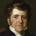 Richard Bright, the father of nephrology
Around 6000 years ago, laboratory medicine began with the analysis of human urine as uroscopy, which later became termed urinalysis. The word uroscopy derives from two Greek words: ouron, which means urine and skopeoa, which means to behold, contemplate, examine, inspect. Ancient physicians spoke of urine as a window to the body’s inner workings and reflected different diseases. Hippocrates (460–355 BC) hypothesized that urine was a filtrate of the humors in the body, originating from the blood filtered through the kidneys. In Aphorisms, he described bubbles on the surface of fresh urine as a sign of long-term kidney disease and associated urinary sediment with fever.
By the late 12th century, a French scholar named Gilles de Corbeil taught and classified 20 different types of urine, recording differences in urine sediment and color. De Corbeil also introduced the matula, a glass vessel in which a physician could assess color, consistency, and clarity. In 1630, Nicolas Fabricius de Peiresc, a French astronomer and naturalist, did the first microscopic description of urine crystals as a heap of rhomboidal bricks. Posteriorly, in the early-mid 1800s, Richard Bright, an English physician, pioneered the field of kidney research leading him to be ultimately recognized as the father of nephrology. These few examples illustrate how urinalysis was the first laboratory test developed in the history of medicine, how it has been persistently used for several thousand years, and how it continues to be a formidable and cost-effective tool to obtain crucial information for diagnostic purposes.
Major improvement in the test-strip technology has been made in recent years. Although dry chemistry technology for urinary test strips has made limited progress, advances in electronic detection have considerably improved the analytical sensitivity of test-strip readers over the years. Not only highly sensitive, test strips are being introduced, but also now, one can find strips, which give quantitative results for urinary proteins. The financial aspect is also of great importance, especially in the Third World and the developing countries; inexpensive test strips for various diagnostic reasons, such as the diagnosis of diabetes form urine sample, are available. Test-strip method also shows promising results in antibiotic susceptibility tests; if optimum diagnostic requirement is reached, it can reduce the test time significantly from 2 to 3 days to few hours.
Urine microscopy is one of the most important diagnostic methods for UTIs and other kidney diseases. In manual microscopy, several steps, such as centrifugation, decantation, and re-suspension, led to cellular lysis and loss. Progress in informatics and computer technology has enabled the development of automated microscopy based on pattern recognition. Over the past two decades, a number of manufacturers have marketed such instruments. Many automated analyzers are now available in the market with different kinds of technologies, such as laminar flow digital imaging technology and pattern recognition technology.
The proteomic method Matrix-assisted laser desorption ionization-time-of-flight mass spectrometry (MALDI-TOF MS) for identification of microorganisms directly from culture coupled with Gram stain has given a new direction, saved considerable amount of time in diagnosis of UTI, and contributed greatly in the field of clinical microbiology in general. It can identify different pathogens accurately and significantly in a short time. The utilization of this technology for the diagnosis of UTIs and furthermore in preforming antibiotic susceptibility tests to decrease the testing time from days to as fast as 2 hours can open doors wide. However, it is unclear whether MALDI-TOF MS can meet the demands of UTI diagnosis, given the need for screening to improve the yield of positive samples. For direct analysis of urine, initial sample preparation steps are necessary to remove cellular debris, WBCs, and mucus, and to collect bacteria.
Adoption of automation in sediment analysis has paved the way for significant progress in urinalysis. These systems reduce human intervention, making it possible to standardize results, and streamline workflow and increase laboratory productivity. Integration of AI with digital bright-field microscopy in automated sediment analyzers provides clear, high-quality images mimicking visual microscopy. These systems are enabled to recognize thousands of known sediment elements. This considerably reduces repetitions, while improving accuracy and reliability of sediment particle detection and differentiation. Additionally, when an atypical cell appears on the screen, the pathologist can check the accuracy of the automatic identification and further evaluate to confirm the sub-types.
Flow cytometry is now being introduced as a reliable method for fast diagnosis of UTIs by counting the bacteria in the urine specimen. Urine particle flow cytometers have improved count precision and accuracy compared with visual microscopy and offer significant labor reduction. With the improved counting precision over visual counting methods, highly accurate positive results can be obtained by this method. Detection of bacteriuria can be achieved with clinical standards using flow cytometry technology.
Smartphone technologies have improved the quality of life in countless fronts, and it has large potential for applications in the medical field. Point-of-care testing (POCT) has been gaining attention for its wide applications in public health. Ranging from blood glucose measurement to complex immunological assays, POCT is simple to use even for end users with little technical expertise. This characteristic makes it particularly suitable for disease detection, quality of care, or patient-compliance monitoring. Although, advancements in semiconductor technology have helped create transformative consumer devices, but so far, have limited impact on the medical world. One of the leading diagnostic tools of POCT analysis is strip-based colorimetric diagnostic assays. When samples, such as urine, blood, or other body fluids, are deposited on a test strip, signals are obtained in the form of colors. With POCT attracting much attention in recent years, smartphone solutions can be a valuable tool in this regard. It can, for example, increase the compliance of populations with screening programs by offering easy and fast screening method. Studies exploring the possibility of establishing a smartphone-based diagnostic platform for rapid detection of zika, chikungunya, and dengue viruses showed valuable prospective. Several other smartphone applications utilizing urinalysis for various diagnostic reasons had been tested, and it shows promising future prospective which can greatly help both medical practitioners and patients alike.
The introduction of POCT allows for more timely diagnosis and rapid clinical decisions toward targeted treatment. Urine dipsticks are a prominently used example of a commercially produced POCT tool for rapid urinary infection testing. There is, however, still room to consider and develop a urine-sampling POCT device that is less costly and more user-friendly. Several groups have developed the concept of a cotton-based smart diaper. Cotton, a natural material, provides excellent flexibility and biocompatibility for detection devices. Above all, there has been an urgent need to integrate urine sampling, testing, and analysis to build a convenient UTI screening solution that would benefit specific, at-risk populations; especially those who have difficulty clearly describing their symptoms. Novel advances could be made to improve the efficiency of UTI screening by using POCT techniques that simultaneously integrate urine collection, screening, and analysis.
Over the past two decades, automated urinalysis has undergone remarkable technical progress. Both microscopy- and flow cytometric-based instruments generate reliable results that are clinically useful, and automated test-strip reading provides added value. Additional integration of existing technologies may further reduce turn-around times. The merging of test-strip technologies with smartphone technologies can lead to tremendous changes in healthcare system and can deeply integrate POCT into the health system. Furthermore, advances in mass spectrometry can lead to great achievement not only in the diagnostic field by providing a much faster and accurate results, but it can also contribute greatly in the medical and biomedical research fields.


