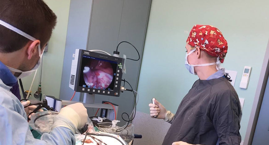
Endoscopy Equipment
Novel techniques are opening up new vistas

Recent developments will enrich the diagnostic toolbox for endoscopists, enhance diagnostic efficiency, and reduce the rate of missed diagnosis and misdiagnosis.
Developments in optical imaging technology continue to promote the revolution of endoscopy, used to examine the interior of a hollow organ or cavity of the human body. Currently, electronic chromoendoscopic techniques such as narrow-band imaging (NBI), linked color imaging, and i-scan provide wide-field high-contrast video images to help endoscopists find mucosal abnormalities especially precancers. However, comparing with traditional histopathologic biopsy, electronic chromoendoscopy lacks the capacity to provide more detailed morphologic information about tissue and cell for accurately confirming or staging cancer. Furthermore, although existing commercial endomicroscopy including endocytoscopy or probe-based confocal laser endomicroscopy can image at cellular information, their detection range is limited to the surface of the mucosa (< 1 mm in depth) which may cause the missed diagnosis of the hidden lesions in deep tissue.
Many studies have been conducted in numerous research centers on novel endoscopic diagnostic technologies to provide more accurate diagnostic information. The advent of these technologies in medical trial research, such as photoacoustic endoscopy (PAE), Raman spectroscopy (RS), two-photon excited fluorescence (TPEF) imaging has opened a new era and created tremendous opportunities for the enhanced identification and biochemical characterization of diseases.
These modalities also have the potential to allow non-invasive in vivo optical biopsy which differentiates areas of similar clinical characteristics, hence challenging the ex vivo histology which is the only way for definitive cancer diagnosis. In addition, these endoscopic techniques own unprecedented temporal-spatial resolution of imaging with innovative mechanisms such as photoacoustics, optical coherent tomography, and multi-photo effect. By utilizing these novel tissue–photon interacting mechanisms, these techniques can guide biopsies by accessing the subtle mucosal/submucosal abnormalities with higher contrast, better resolution, and penetration depth. Hence, they cut down the number of biopsy times, costs, and risks for patients.
However, most of these technologies are not mature, so they have been neither officially launched into the market nor put into clinical use in the hospital so far. Thus, well-organized large-scale animal experiments and human clinical trials should be executed to further validate and standardize these techniques before approval of the clinical application.
Currently, endoscopic ultrasound (EUS) is the only clinically available tomographic endoscopic tool used to diagnose various diseases. EUS cannot receive high-resolution vasculature information of the suspicious region, owing to the limited detectable depth and the operating mechanism. Although, photoacoustic spectroscopy and simple imaging were developed in the 1970s, photoacoustic imaging only recently became famous in biomedical research.
 Dr Nikhil Arbatti
Consultant Spine Surgeon,
Minimally Invasive Spine Surgery Specialist
“With the pandemic in 2020, the elderly with spinal ailments have been affected the most. Many of them were restricted to home, instances of falls leading to spinal fractures or worsening of spinal problems has increased. Many of them now want to get there spinal problems treated surgically, but due to the fear of long surgeries and extended stay in hospitals, they are either postponing or avoiding spinal surgeries. Endoscopic and minimally invasive spine surgeries go a long way in solving these problems. With minimal incisions, practically no blood loss and minimal pain post-surgery, the patients are mobilized post-surgery immediately and can be discharged the same day. Thus, the elderly population is the one which is benefitted the most. It is estimated that endoscopic/minimally spinal surgeries will grow to almost 50 percent of the spinal surgeries happening today by 2030″
The advent of photoacoustic endoscopy (PAE) aims to overcome the EUS’s limitations by adapting the photoacoustic effect. Photoacoustics can be described as a laser-induced ultrasound. Short light pulses (nanosecond range) are absorbed by tissue absorbers accordingly (such as endogenous contrast: hemoglobin, melanin, and exogenous contrast: methylene blue). Then a transient temperature increase is generated, resulting in local thermoelastic expansion effect, which gives rise to ultrasonic waves. After that, the ultrasonic wave signal is recorded by nearby detectors.
Compared with NBI, CLE, and OCT, PAE provides functional optical contrast with high spatial resolution while maintaining the benefits of greater penetration depth of traditional ultrasound endoscopy. Particularly, PAE embodies photoacoustic tomography (PAT), which is cross-sectional or three-dimensional imaging using the photoacoustic effect. Thus, PAE can offer in vivo 3D volumetric images of biological tissues with high spatial resolution and stark tissue-optical contrast. Thus, characteristics of the targeted area, like architectural vascular features and dynamic changes of tumors, can be clearly observed from the 3D images.
To conclude, PAE has great potential for in vivo endoscopic applications, such as precancer detection, accurate diagnosis of submucosal abnormalities, and in situ characterization of diseased tissues. Endogenous or exogenous contrast agents may improve the ability of endoscopic imaging, resulting in the earlier and more accurate detection of malignant and premalignant lesions. The wealth of experience gained in preclinical studies as well as recent technological progress provides a solid foundation for photoacoustics to advance into mainstream use.
Nonetheless, further refinement of photoacoustic imaging is necessary for broader dissemination, including stable laser systems, user-friendly imaging devices, algorithms, and compatible software that can robustly analyze imaging data to extract diagnostically relevant quantity. Moreover, the most apparent limitation remains the penetration depth of light. The near-infrared light is not sufficient to visualize targets beyond a few centimeters under the tissue surface. Additionally, the imaging speed of PAE systems is relatively low, so there is still a long way to implement real-time detection.
Multi-photon imaging is a promising technique for label-free imaging of biological samples. The multi-photon effect refers to the phenomenon that a molecule simultaneously absorbs or scatters two or more nonlinear photons with the same frequency, relaxes to the ground state after reaching the high energy state, and emits shorter wavelengths which can be used for biomedical imaging. Compared to single-photon imaging, multi-photon imaging uses a near-IR light source that is able to create sufficient peak energy to produce multi-photon effect while keeping average energy low enough to minimize specimen damage. There are several nonlinear imaging modalities, such as two-photon excited fluorescence (TPEF), three-photon excited fluorescence (3PEF), second-harmonic generation (SHG), and third-harmonic generation (THG) imaging. They can provide valuable information about the biological samples’ intrinsic properties, which can be employed in diagnosis applications.
It is challenging to acquire high-resolution images from tissue in the deep layer, for the high degree scattering caused by discontinuous refractive index and heterogeneous constituents. Despite that, the near-IR owns deeper penetration depth and causes less damaging than shorter wavelengths. Thus, the infrared light can be selected as the excitation light source for multi-photon imaging, which effectively reduces the scattering of excitation light and gets a high signal-to-noise ratio by avoiding the interference of single-photon auto-fluorescence.
Furthermore, no pinhole is required to prevent out-of-focus fluorescence from reaching the detector in MPE, because excitation occurs only at the excitation spot, which results in the optical sectioning inherent to nonlinear imaging techniques. MPE owns the advantages of strong penetrability, high resolution, low phototoxicity, and no need to stain the tissue. MPE can ex vivo or in vivo image and detect early invasion of cancer in reconstructed 3D photos as well as 2D views. The critical challenge of realizing MPE is how to miniaturize the system into a compact and lightweight distal probe while keeping the general performance.
Raman Spectroscopy (RS) is an exciting analytical optical spectroscopic technique capable of probe vibrational and rotational modes of endogenous biomolecules of tissue or cells intrinsically. Photons in the incident laser light interact with the tissue and undergo inelastic collisions with molecules, resulting in an exchange of energy and a change in frequency. Moreover, in the Raman spectrum, the intensity of the peak of shifted light is directly proportional to the concentration of the sum of molecular constituents giving rise to that peak. Thus, the Raman spectrum is a direct function of the molecular composition of the targeted region within the tissue, giving us a complex molecular fingerprint. Most biological molecules have distinct vibrational energies in their molecular bonds. Thus, they have their specific fingerprint. In addition, the Raman shift caused by frequency change is specific to the species of the molecule. The shifts in wavelengths are expressed as wave numbers, and they are independent of the wavelength of excitation, which means that the energy shift is constant for particular molecular bonds.
By measuring the molecular specific inelastic scattering of light, RS enables the explanation of a tissue’s biochemical fingerprint. Primarily, RS requires a monochromatic laser to illuminate the tissue sample. Subsequently, the intensity and wavelength of the scattered light are collected and analyzed. Therefore, RS can detect subtle biochemical and molecular changes that are vital in the discrimination of tissue samples, which makes RS can become a potential diagnostic tool. However, the inherent weakness of Raman signals is one major limitation of RS. Thus, endoscopic RS systems need high laser powers and ultrasensitive detectors, which are enough to merit this technique with clinical value. Moreover, the sensitivity can also be increased with the Raman signals enhanced by ten orders of magnitude by a phenomenon known as Surface Enhanced Raman Spectroscopy (SERS).
RS has excelled in the early detection of precancer and cancer in the interior of various organs with high diagnostic specificity. Several probes are currently undergoing construction, and engineering tolerance levels are being carefully defined before a clinical trial. Further evaluations of the clinical merits of Raman spectroscopy for prospective prediction of precancer and cancer in vivo are expected.
Before any novel endoscopic optical diagnostic technology is successfully put into clinical use, it is imperative that ex vivo/in vivo preclinical animal experiments and ex vivo human trials be conducted, which are followed by rigorous in vivo human clinical trials to further verify the effectiveness and safety of these techniques.
Furthermore, these emerging novel endoscopic optical diagnostic techniques will continue to develop with the cooperation of multi-disciplinary to improve the diagnostic capabilities of detection, characterization, and confirmation in the near future. To obtain certification of the supervisory authorities on medical devices in different regions all over the world, such as Food and Drug Administration (FDA) and European Conformity (CE), current efforts are directed to implement and validate these technologies for modern endoscopy before general clinical practice in hospitals. However, these emerging diagnostic devices have not obtained official approval of clinical use and vary across maturities. Some challenges lie in the results inconsistency, cost-effectiveness, time-efficiency, etc., which require further researches and developments to achieve a promising era of endoscopy.


