
MB Stories
Recent advancements in MRI technology – Improving accuracy, speed, and safety
Recent innovations offer new possibilities to expand the use of MRI.

Innovation in radiology and imaging has been an area of increased focus in the medical industry over the past decade, with new technologies and visualization advancements emerging at a rapid pace. Artificial Intelligence (AI) implementation and advancement in imaging technologies have been crucial to developing more accurate results, thereby improving diagnoses and patient treatment options.
Innovations in medical imaging are clearly a game changer but the direct impact on patient and economic outcomes is quite often understated. Now AI algorithms are being used to analyze millions of medical images in real time, enabling the detection of early-stage diseases and conditions. For example, it is being used to improve accuracies of mammograms, a crucial tool for detecting breast cancer, as well as advanced analyses of MRIs to help detect and treat prostate cancers. These algorithms analyze images faster and more accurately than human radiologists, reducing the sole reliance on manual image interpretation and minimizing risks of missed diagnoses. Advanced MRI techniques have been in regular development, making the procedure faster, safer, and more efficient. New techniques, such as compressed sensing MRI allow for faster scans, with fewer scans required to produce the same level of detail. This can be especially beneficial for patients with mobility issues, or children who struggle to remain still during scans. Whole-body magnetic resonance imaging (WBMRI), for example, is the most sensitive imaging test, allowing for earlier detection and treatment of multiple myeloma, directly contributing to improved long-term health and economic benefits.
Additionally, computed tomography scans, widely used to produce detailed images of internal organs and tissues, can be associated with significant radiation exposure. New and improved technologies include dual-energy CT scans, which reduce radiation doses, while providing high quality images. These scans use two different types of radiation to produce images, enabling radiologists to differentiate between healthy and diseased tissues more accurately. The introduction of teleradiology has become a focus of digital transformation in healthcare systems, which are continuously facing challenges in meeting the demands for medical imaging services, particularly in rural and remote areas. Teleradiology is helping to bridge the gap between patients and radiologists, providing improved access to imaging services and reducing wait times, resulting in much faster diagnoses and reduced burden on practitioners.
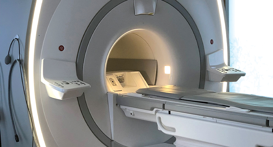
Technology trends
In recent years, several advancements have been made in medical MRI technology.
Some of the latest developments in MRI technology to see what will be shaping the conversation around magnetic resonance imaging for years to come:
Faster scanning. Advances in MRI technology have allowed for faster scanning times, reducing the amount of time patients need to spend in the scanner. This not only makes the experience more comfortable for patients, but also allows for a quicker turnaround time in obtaining results. This can be especially important in emergency situations or when patients have trouble remaining still during the scan.
Improved image quality. Improved hardware and software have led to better resolution and contrast in MRI images, providing a clearer and more detailed picture of what is happening inside the body. This can help medical professionals make more accurate diagnoses and develop more effective treatment plans.
3-D MRI scanning. The three-dimensional (3-D) MRI scanning software allows MRI machines to upgrade from a traditional two-dimensional system, facilitating a more transparent and comprehensive patient-screening procedure.
Conventional devices perform scans in sections or slices and from top to bottom. This computing technology puts together these captured images to create a 3-D representation that is more useful in the diagnosis, monitoring, and pre-surgical procedures, complementary to other laboratory test results.
Open MRI machine. Some patients are not comfortable lying down and getting the feeling of going through a tunnel, which can be challenging for those with claustrophobia. With technology, manufacturers have developed an open MRI, making the procedure less terrifying for persons with anxiety. This advanced device eliminates the enclosures in the front and back sections, helping ease fear and anxiety in most people.
An open MRI is also more inclusive, allowing obese people to go through the exam without the fear of getting stuck inside. And because it can be done while the patient is either standing or sitting, it is more comfortable than the traditional method.
MRI 0.55T is here. For years, the conventional wisdom held that, in order to get a high-quality MRI exam, you need a field strength of 1.5T or above. However, that conventional wisdom has been proven wrong with the advent of .55T MRI. Known as High-V MRI, systems with this field strength take the power of digitization and deliberately apply it to a new field strength of .55T with inherent clinical benefits.
High-V MRI does this in a few different ways:
- Improved implant imaging. Imaging metal implants has historically been difficult with conventional MRI systems because of the artifacts that result. High-V MRI offers intrinsic physical advantages that result in reduced metal distortions, leading to strongly improved diagnostic capabilities for implant imaging.
- Reduced susceptibility challenges. Susceptibility artifacts are a well-known phenomenon in MRI. One prominent example is an air-tissue interface that occurs at the sinuses and orbits. The unique field strength of High-V MRI offers physical advantages that inherently reduce these susceptibility artifacts. This leads to reduced geometric distortions in diffusion imaging, resulting in improved diagnostic quality.
- Pulmonary imaging opportunities. Pulmonary imaging has been notoriously difficult with conventional MRI as the air-tissue interfaces result in a fast signal decay. These challenges scale with magnetic field strength, which makes High-V MRI the perfect opportunity for pulmonary imaging. High-V MRI provides possibilities to expand the reach of your pulmonary MRI capabilities.
Weight-bearing MRI methods. Like open MRI tools, some machines allow patients to perform weight-bearing positions, such as sitting down while being scanned. These machines suit patients with specific conditions that make lying down uncomfortable, such as those with compression and disc herniations. Because the patient does not have to lie flat on their stomach or back, this procedure is ideal for patients with spinal and sports injuries, or musculoskeletal disorders that make every movement unbearable.
Surgical MRI machines. With technology, MRI procedures are no longer used for diagnostic purposes only. With intraoperative magnetic resonance imaging (iMRI), surgeons can use the machine inside the operating room to make the procedure more accurate. Surgical doctors can use these advanced systems to view high-resolution images of the surgery site, helping them target the problematic area with better precision.
Patient-friendly MRI devices. Besides an open MRI, technology has enabled other integrations to make the procedure less likely to elicit fear and anxiety from patients. For instance, some MRI devices allow patients to relax while enjoying an integrated entertainment system that provides the best audiovisual experience. Instead of being flooded with thoughts about what could go wrong, individuals undergoing the exam can sit back and relax.
Silent MRI scanning is another patient-friendly technology embedded in some machines. It cancels some noises that can elicit annoyance and irritation to some people going through the procedure. Patients will have to wear noise-canceling ear devices to make them feel more comfortable.
Enhanced safety. Newer MRI machines use stronger magnets and advanced shielding to reduce the risks associated with the magnetic field and radio waves used in the scanning process. This makes MRI a safer option for patients, especially those who have metal implants or other conditions that may pose a risk during the scan.
Increased accessibility. Portable MRI machines and open MRI machines have been developed, providing increased access to MRI scans for patients who may have difficulty getting into a traditional MRI machine. This includes patients with mobility issues, anxiety about enclosed spaces, or larger body sizes.
Emerging applications. MRI technology is being used in new and innovative ways, including functional MRI (fMRI) to study brain activity, diffusion MRI to map the structure of white matter in the brain, and MR angiography to visualize blood vessels. These advancements are helping to expand the capabilities of MRI and improve patient care.
Pulmonary MRI. Getting a view of the lungs and what is inside them traditionally requires other medical imaging types, such as computed tomography (CT) scan. Pulmonary MRI was highly challenging in the past because the machine requires low levels of hydrogen atoms while our lungs are filled with oxygen air. As a result, radio wave signals could not stay stable enough to get an accurate image. A manufacturer then created a machine that facilitates lung tissue viewing through ultrashort echo time (UTE) technology that stabilizes the signals.
Cost-effective and simple cardiac MRI. Because of its complex nature and the hefty costs involved, only a small number of cardiac MRIs are performed even in advanced countries like the United States. Most medical practitioners still rely on CT scans to take images of the heart. Thus, an electronics manufacturing giant introduced a new technology to make cardiac MRI procedures less costly and more straightforward.
The software, which works with specific machines, automates images taken from a 3-D chest scan. It enables devices to capture a complete pattern of one cardiac movement, including blood-flow conditions. This technology also reduces the testing time from over an hour to only 10 minutes.
Multi-contrast imaging. Even if MRI scanning is considered one of the most advanced medical imaging methods, there are areas for improvement. For instance, machines of the past were not able to provide the most detailed images at all times. As a result, a rescan will be ordered if the doctor cannot interpret the images well. With multi-contrasting imaging software, MRI machines can take eight contrast settings in one examination. There is no need to order a rescan because medical practitioners can look at several results in different contrast settings. By adjusting contrast settings, doctors can better view what is inside your body to come up with better diagnoses and treatment plans.
Software-based developments in MRI. The advancements of the MRI system are not only confined to the equipment side. There are developments in the software edge of the system as well. Siemens multi-slice application is a software developed to obtain MR images simultaneously rather than sequentially, thus enabling time-saving while 2-D imaging. GE HealthCare has developed MAGiC (MAGnetic imaging Compilation) software. This software is the first multi-contrast software that permits eight contrasts in a single acquisition. This increases the efficiency of diagnosis with MRI.
Outlook
Overall, these advancements in medical MRI technology are improving the accuracy, speed, and safety of scans, and providing medical professionals with more information to make better decisions about patient care.
As technology continues to evolve, it remains to be seen which advancements in the MRI system will be introduced in the coming days. Nevertheless, it cannot be denied that these state-of-the-art tools are already helping make the medical practitioners’ lives easier and the patients’ better.


