MRI Equipment
Scanning the MRI horizon
MRI has been perceived as a futuristic imaging technology for the past few decades. That might be about to change with the advent of new MRI technologies released with a concentration in new ways to speed upimage acquisition.
 The technical capabilities of current MRI systems have been driven largely by advances in computers, material science, engineering, and physics. Although some extraordinary feats, such as single-atom imaging and ultrahigh-field anatomical imaging, have received considerable media attention, there are a myriad of incremental advances that have improved clinical imaging.
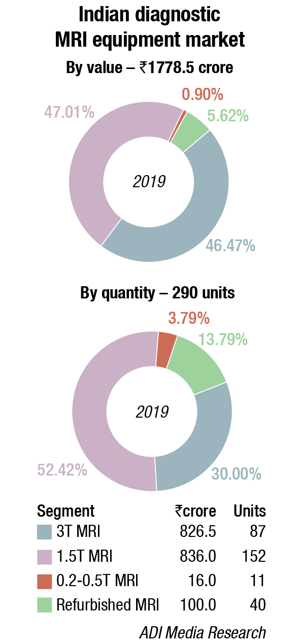
Better image quality and higher spatial resolution of today’s MRI are largely due to advances in MRI pulse sequences and hardware, including higher field strengths, improved multichannel coils, stronger gradients, and more homogenous magnets. Whereas it was common to image at 0.5 and 1.5 T several years ago, many magnets currently operate at 3 T. Today, coils that collect the body’s MRI signatures are typically phased array, flexible printed, or blanket coils rather than the bulky cage coils used a decade ago. Beyond improving image quality, these advances have enabled functional MRI (the combination of morphological data with biological information) of the brain, vascular mapping, and real-time cardiac imaging. Engineering advances have also led to development of wide-bore and open magnets, allowing interventional procedures and surgeries and accommodating claustrophobic patients.
Faster imaging has become a clinical reality, although much work remains to be done in this area. Increasing imaging speed improves patient comfort and throughput, decreases cost, reduces motion artifacts, and enables imaging of joints and muscle function, such as in the heart. Newer approaches to achieve faster imaging include undersampling of K-space (partial Fourier reconstruction and parallel imaging), compressed sensing, and non-Cartesian sampling with constantly improving reconstruction algorithms.
Computational analysis and artificial intelligence (AI) play increasingly important roles in all aspects of MRI acquisition and data processing. For example, AI-based image reconstruction greatly reduces the time required for image reconstruction, producing images of comparable quality while providing the ability to reconstruct large datasets in near real time. In addition, the increasing availability of AI has laid the foundation for automated image post-processing, including segmentation and volumetric analysis. Volumetric analysis is particularly useful in imaging Alzheimer’s dementia and medial temporal lobe quantification, although predominantly used in a research setting at present. Last, AI can be used for decision support to create automated reports, flag imaging abnormalities, or classify organ structures as normal or abnormal.
 Dr Kanchan Sonone
Consultant and Lab Director, Wockhardt Hospital
The next decade of clinical MRI
“When talking about biggest discoveries, medical inventions are no exceptions, especially when talking about crucial and extraordinary inventions such as MRI scanning. The benefits of MRI scanning convenes beyond the need for single diagnosis or finding the problem, as one can use the features and benefits of MRI in treatment and prevention.”
Although clinical challenges abound for MRI, the opportunities are equally expansive. Looking forward, the most pressing clinical needs are shortening MRI acquisition times, optimizing image quality and content, automating analyses, perfecting fusion imaging, and enabling whole-body imaging.
Deep learning (DL) algorithms are likely to play a central role in image acquisition (sub-Nyquist sampling strategies using DL), reconstruction, and automated image post-processing. More seamless integration of imaging results into electronic medical records will be essential. Currently, this remains cumbersome because the different imaging technologies and platforms are not optimized to interact with and learn from each other.
Of the many potential clinical advances likely to result from expanding imaging technologies, several hold notable potential. A key interest in neurologic imaging is to translate emerging ultrahigh-field MRI (>3 T), which increases the signal-to-noise ratio, into clinical practice. This will allow better anatomic and ultrastructural imaging and will likely open new doors to disease characterization. Although a few such ultrahigh-field systems are operational in clinical research settings, practical challenges will need to be overcome for the approach to be adopted more broadly.
Advanced thoracic imaging will require the development of image acquisition methods during quiet breathing rather than the current approach of breath holding, which often is difficult for patients.
While acknowledging the advances made in decreasing scan time and optimizing image acquisition over the past decade, there remains considerable potential for improving the patient experience in the MRI suite. At present, advanced imaging techniques, such as functional and cardiovascular MR, require protracted scan times (sometimes up to or more than an hour). Consequently, patients often become uncomfortable during the examination, and motion artifacts remain a considerable problem. Although retrospective motion correction algorithms are useful, adaptive dynamic imaging, including prospective motion correction currently in development, is expected to expand the applications of cardiovascular and functional MRI. Within the field of cardiovascular imaging, efforts are being made to combine these prospective motion correction algorithms with free breathing techniques to further enhance the patient experience. Previously, complex sequence acquisition required electrocardiogram gating, breath-hold sequences, and respiratory gating to achieve artifact-free images. New efforts are emerging on MR multitasking, which continuously collects geometric data and resolves for these artifacts. Undoubtedly, the secondary gains from this progress will include enhanced image quality, shorter scan times, and improved throughput.
Realizing the full potential
MRI has been perceived as a futuristic imaging technology for the past few decades. However, its expense, complexity, long imaging exam protocols, and long post-processing times have presented a barrier to wider adoption.
The advances in MRI technologies described have not been realized without growing pains, and a considerable amount
of work must be done to improve them further. The aspirations of the field are ambitious and will require a community of basic and translational scientists, as well as physicians, to achieve them. Such efforts have been catalytic in the past, as evidenced by the expeditious development of MRI in the past decade. Many new applications will necessitate prospective clinical trials and cost-effectiveness analyses so that emerging techniques can become reimbursable. Commensurate advances in the allied fields of AI and MR physics will be necessary to facilitate all of these improvements. Collaborative efforts and public-private partnerships will be essential to driving these technologies and realizing the full potential of MRI in the years to come.


