
MedTech
The evolving role Of technology in MRI
New MRI technologies released in the last few years are changing the fortunes of this sector.

Magnetic resonance imaging (MRI) for cardiac assessment provides a radiation-free alternative to other commonly used modalities like computed tomography (CT) and single-photon emission computed tomography (SPECT). Cardiac MRI (CMR) offers greater contrast and image clarity than CT, does not require the use of a contrast agent, and allows radiation-free perfusion imaging. The technology does, however, present challenges unseen with other modalities. It remains the most expensive of all imaging exams, largely thanks to the length and complications of the scan itself. Furthermore, the use of magnets has traditionally made MRI inaccessible for patients with implanted medical devices. As a result, CT and SPECT have remained the gold standard for cardiac imaging assessment.
While these modalities will continue to serve important roles, MRI technological advancements are providing new opportunities for cardiac MRI to expand. CMR has a distinct advantage over echocardiography and nuclear scintigraphy in terms of fewer artifacts and unlimited acquisition windows. It is noninvasive, lacks ionizing radiation, and uses benign paramagnetic contrast agents without nephrotoxicity. It has positioned itself to become a one-stop diagnostic shop, offering extensive results during a single examination, currently only available through a combination of multiple, separate, diagnostic procedures.
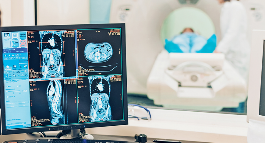
Perhaps no technology has generated more buzz in radiology recently than artificial intelligence (AI). The technology is a potential game-changer in the space, essentially giving radiologists a second set of eyes for diagnosis and image analysis.
Now, new research sheds light on just far industry has come in terms of the development of AI and machine learning (ML) programs. According to a recent study published in the journal, Circulation: Cardiovascular Imaging, analysis of cardiac MRI scans using automated ML can be performed significantly faster and with comparable accuracy to human interpretation by trained cardiologists. The question that arises is: will it be able to shed human input?
AI, a major tool for cardiologists
It generally takes about 13 minutes for a trained physician or cardiologist to interpret a cardiac MRI. But with the advent of AI and ML algorithms, a scan can be analyzed with similar accuracy in approximately 4 seconds – nearly 186 times faster! Cardiac MRI scans help cardiologists make preoperative decisions for surgical planning by providing important measurements of heart structure and function. But they may also help to make decisions related to the timing of cardiac surgery, implantation of defibrillators, and continuing or discontinuing infusions of cardiotoxic chemotherapy for cancer patients. Such clinical decisions ultimately rely on accurate and precise measurements from cardiac MRI scans.
Acceleration of data gleaned from such MRI scans might also help to improve the speed of clinical decision-making and subsequent health outcomes, ultimately affecting care from a population health perspective.
A study was performed in the UK, where approximately 150,000 cardiac MRI scans are performed annually. Extrapolating the number of scans performed per year, the investigators calculated that using AI to interpret scans could potentially result in saving 54 clinician-days annually at each health facility. For their study, researchers developed and trained a neural network to read the cardiac MRI scans of nearly 600 patients. They found that there was no significant difference in accuracy when the machine learning algorithm was tested for precision compared to an expert and trainee on 110 separate patients from multiple centers.
Cardiovascular MRI offers unparalleled image quality for assessing heart structure and function; however, current manual analysis remains basic and outdated. Automated ML techniques offer the potential to change this and radically improve efficiency, and experts look forward to further research that could validate its superiority to human analysis.
Machine learning will impact how cardiologists read cardiac MRI studies in their day-to-day work and how they train future generations of physicians. Adoption of novel tools that increase precision and efficiency are useful not only for individual patient care but also in performing larger-scale research studies.
Having said that, an ML algorithm does not perform better compared with human interpretation. The industry is not at a point where machines can go unchecked and unsupervised after they deliver an interpretation.
Clinicians still need to provide oversight and verify readings to assure patient safety and quality of care. While a reduction in clinical hours is certainly an added benefit to such technology, one must first ensure that there is no patient harm in larger-scale studies.
However, the potential utility of AI as a transformative technique in increasing speed, without sacrificing accuracy and precision for the interpretation of complex imaging scans for patients with structural heart disease, cannot be undermined.
Limitations of machine learning. ML, a subset of AI, has allowed the field of cardiology to escape the confines of conventional inquiry, and embark on a new realm of multi-dimensional information occurring in real-time leading to data discovery-driven research. ML is not a distant concept on the horizon but an inevitable necessity in the evolutionary line of cardiovascular imaging and clinical care, especially in CMR.
ML will become an inevitable necessity as the number of medical apps, wearable devices, and miniaturized devices continue to grow and prosper. For successful integration into the clinical environment, some issues need to be addressed. ML algorithms require extensive exposure to large datasets to gain accuracy, and obtaining such datasets can be difficult. Data sharing among institutions can be difficult and laborious because of multiple institutional review boards, and datasets ideally should be publicly available.
A universal standard is required for data standardization. Although digital imaging and communications in medicine (DICOM) and picture archiving and communication systems (PACS) are valuable for imaging data, there are differences in these programs between centers. There are many different classifications, protocols, and acquisition protocols among various institutions, and imaging and clinical data exist on separate user interfaces. As data become larger and more complex, manual data entry will become difficult. If newer software can successfully integrate clinical and imaging information, it can facilitate the expansion and utilization of ML in various academic centers.
Outlook
MRI has been perceived as a futuristic imaging technology for the past few decades. However, its expense, complexity, long imaging exam protocols, and long post-processing times have presented a barrier to wider adoption.
That might be about to change with the advent of new MRI technologies released with a concentration in new ways to speed up the image acquisition. This includes techniques like compressed sensing, parallel imaging, and echo sharing. AI too is playing a significant role in MRI automation to speed workflow and quantification, and it is expected to become mainstream in the next few years.
Second Opinion
 Dr Aditya Gupta
Neurosurgeon,
Artemis Hospital
MRI, making things clear!
MRI, which stands for magnetic resonance imaging, is a technology that has revolutionized many aspects of modern medicine.
To put it simply, it involves exciting the hydrogen atoms in the tissue by delivering magnetic energy pulses, and then measuring the energy emitted when these atoms return to baseline energy. Since almost all human tissues have high water content, this hydrogen atom signature delineates practically all tissues at a very high level of spatial detail.
The high-resolution images provide the minutest of details about many organs, which are extremely helpful in making diagnosis. One prime example is neurological disorders.
Special sequences permit measurement of particular molecules in tissues, and certain sequences enable even blood flow in the blood vessels to be quantified. It is no surprise that diseases such like brain and spine tumors, stroke, multiple sclerosis, and infections have become much more treatable once such accurate diagnosis has become possible. An abnormal area as small as 1 mm can be detected by MRI.
MRI-guided PET scans are revolutionizing cancer detection at early stages in practically any part of the body.
Another revolutionary advance is the use of MRI-guided focused ultrasound (MRIgFUS) to treat movement disorders by noninvasive lesioning in the brain, and also to deliver chemotherapy agents by modifying the blood-brain barrier.
MRI scanners are now available in practically every small town in India. This technology is clearly serving an important role in healthcare, which is only poised to increase in the near future.


