


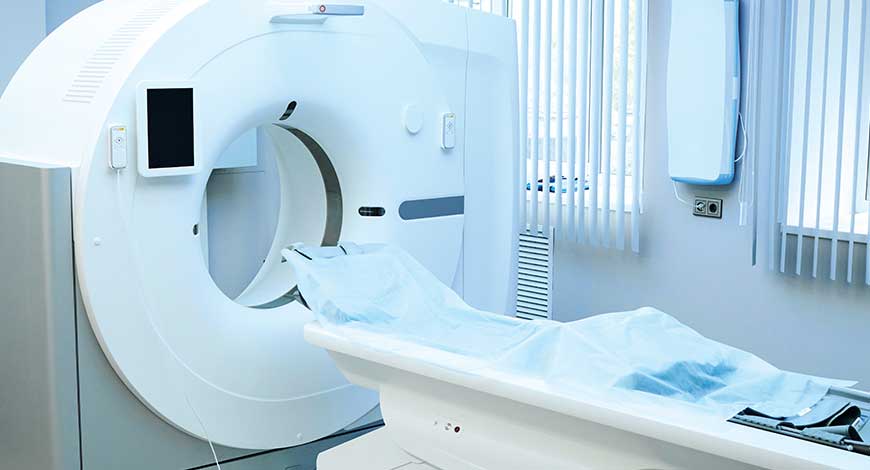
CT Scanners
CT scanners | Continue to be a cornerstone of modern medical diagnostics
With advancements in resolution, speed, radiation dose reduction, AI integration and personalized medicine, CT scans will continue to be a cornerstone of modern medical diagnostics.

Computer tomography has proved fundamental in image evaluation throughout the past three decades. By combining rapid scanning with high-quality data sets, multi-detector technology continues to influence practice patterns. This has led to new applications and improved use in conventional applications.
CT scanners have become main choice of investigation after x-ray. CT scanner is taking x-rays across the patient body in 360 degrees. With advancement in technology, the scan time taken to rotate 360 degrees around the patient is reducing, thus enabling CT scanners to image heart. CT coronary angio procedures are increasing and making this a part of preventive healthcare check-up after age of 40 years; lowering the x-ray dose to patients has taken an important step for future developments.
Technological advancements
One of the most striking aspects of modern CT scanners is their speed and accuracy. Advanced multi-slice CT scanners can capture multiple images within seconds, enabling swift and comprehensive assessments. This speed reduces examination times and minimizes patient discomfort by avoiding the need for repeated scans. Moreover, the integration of cutting-edge technologies has equipped CT scanners with enhanced diagnostic prowess, such as dual-energy CT for improved tissue differentiation and spectral imaging for enhanced contrast and tissue characterization.
Some of the technological advancements taking place in today’s market reveal the dedication to enhancing patient care, diminishing radiation exposure, elevating image quality, and expanding the utility of CT across medical specialties.
One of the most anticipated developments in CT technology has been the improvement in resolution and speed. As computational power and software algorithms continue to advance, CT scanners are expected to produce even more detailed images in a shorter amount of time. This benefits patient comfort and enables more precise diagnoses, particularly for conditions that require capturing dynamic processes within the body.
The increased use of CT has generated significant concern regarding the high radiation doses received by patients during CT scans compared to traditional radiography examinations. Many studies have been undertaken on minimizing patient dose and adhering to as low as reasonably achievable (ALARA) principle.
New models are being designed to provide exceptional image quality with significantly lower radiation doses, minimizing the risks associated with frequent or repeated scans. Techniques such as iterative reconstruction algorithms and automatic exposure control, have been developed to minimize radiation dose while maintaining image quality. This trend is expected to continue with a focus on dose optimization.
Advancements in miniaturization and portable technology will likely lead to the development of compact, point-of-care CT scanners. These devices could be used in emergency rooms, ambulances, and even remote locations, providing quick access to diagnostic imaging in critical situations.
Researchers are pioneering approaches to illuminate and radio-sensitize tumors using gold-based nanomedicine. This innovative strategy holds promise for targeted CT imaging and therapy. By leveraging 2DG-bound gold-based nanomedicine, the research seeks to enhance precision and effectiveness in both diagnostic imaging and therapeutic interventions. This not only contributes to the evolving landscape of cancer treatment but also exemplifies the intersection of nanotechnology and medical imaging for improved patient outcomes.
Several centres are still using chest radiography as the imaging technique of choice in infants and preschool children, although CT has shown higher sensitivity and specificity in detecting early abnormalities in both symptomatic and asymptomatic CF (cystic fibrosis-related lung damage) patients.
The sensitivity of chest radiography is poor, and the variability among radiologists in interpreting chest radiography is high, even when combined with adequate scoring systems. Furthermore, CT contributes significantly to clinical decision. A recent study showed that CT is a simple stratification tool that can identify children at increased risk of poor outcomes in later life and is an early indicator of the effectiveness of interventions in very young children who have disease-modifying potential. The use of chest radiography is also debated during pulmonary exacerbation, especially in children with advanced disease . Children with CF and advanced lung disease are at higher risk of pulmonary exacerbation, and on chest radiography it is more difficult to detect any new radiographic change in a lung substrate with diffuse parenchymal abnormality. Similarly, in uncooperative children, those who cannot follow breathing instructions, given the speed of the latest generation of CT scanners, the entire chest can be scanned in less than a second, and also without anaesthesia.
Artificial Intelligence
AI is poised to revolutionize image reconstruction and interpretation, offering the potential to significantly improve diagnostic accuracy and efficiency. This technology has the capacity to transform CT imaging from a mere diagnostic tool into a powerful instrument capable of not only detecting abnormalities but also correlating these features directly with specific diseases. With the integration of AI into CT imaging, traditional methods can be enhanced by automatically identifying changes in organ features indicative of disease.
A groundbreaking study from the University of Gothenburg, Sweden has introduced a novel method capable of extracting comprehensive diagnostic information from brain images obtained through CT, rivaling the detail provided by MRI. Despite CT’s widespread availability and affordability compared to MRI, it has traditionally been deemed less effective in revealing subtle brain structural changes.
This new approach, leveraging AI trained on MRI data, bridges this gap by enhancing the interpretation of CT scans, particularly in diagnosing conditions like dementia. The software developed through deep learning AI demonstrates promising potential to significantly augment diagnostic support, especially in primary care settings. By accurately measuring brain structures and volumes from routine CT scans, the method offers a rapid and reliable means of decision-making support, aiming to reduce false negatives and streamline patient referrals to specialist care.
Notably, the software’s versatility extends beyond dementia diagnosis, with ongoing investigations into its utility for conditions like normal pressure hydrocephalus (NPH), where it shows promise in both diagnosis and treatment monitoring. As development progresses in collaboration with international clinics and companies, this innovative solution holds significant promise for transforming imaging diagnosis and improving patient outcomes in neurological healthcare.
Another development using AI software is early-stage lung cancer detection, an abnormality spotted on a chest CT scan. These lung nodules, which can be as small as a few millimeters, are challenging to identify and may be overlooked. However, AI software has shown promise in aiding radiologists in this task.
Researcher Ward Hendrix from the Diagnostic Image Analysis Group, Netherlands has developed and validated AI software for detecting lung nodules in non-screening CT scans at Radboudumc and Jeroen Bosch Hospital. This collaborative research, initiated by Colin Jacobs and other researchers was published in the journal, Communications Medicine.
The AI software accurately located cancerous and non-cancerous nodules, including those in common blind spots of radiologists. These findings suggest that AI technology is mature enough to detect lung nodules in diverse CT scans and has the potential to assist radiologists in this regard.
Technological advancements are taking place at a rapid pace, revolutionizing various industries including healthcare. But, threats such as cybersecurity vulnerabilities still persist, posing significant challenges to the safety and integrity of critical systems like CT scanners.
Outlook
CT technology holds immense promise for both healthcare providers and patients. With advancements in resolution, speed, radiation dose reduction, AI integration and personalized medicine, CT scans will continue to be a cornerstone of modern medical diagnostics. The shift toward functional and multimodal imaging, along with the development of portable CT scanners, will further extend the reach of this technology. As CT technology evolves, it will undoubtedly contribute to more accurate diagnoses, better treatment outcomes and improved patient care in the years to come.


