


Trends
MRI scans pinpoint root of lingering concussion symptoms
MRI scans recently revealed features that link lingering post-concussion symptoms with a possible source of vestibular dysfunction in the brain.

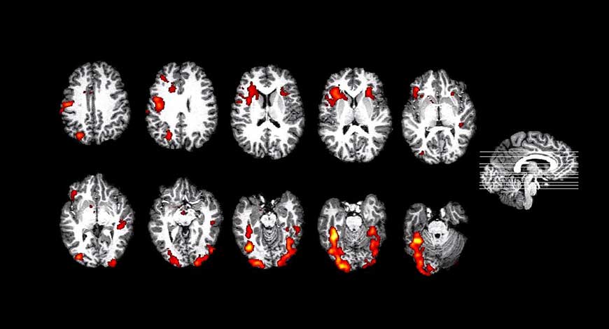
A new study published in the Journal of Neurotrauma analyzed imaging from 42 athletes who underwent multiple MRI sequences to pinpoint differences in the brains of those who had sustained head trauma and those who had not. Researchers found that more than half of the symptomatic athletes who reported head trauma showed signs of injury to the inferior vestibular nerve on imaging. These findings could be to blame for why individuals who have sustained a concussion continue to have symptoms like dizziness, headaches, difficulty focusing and issues with balance long after their initial injury, the researchers suggested.
“While symptoms typically gradually subside within 7-14 days, some athletes show prolonged persisting symptoms, and some never recover fully,” first author on the study Anna Gard, MD, at Skåne University Hospital, and co-authors stated. “Vestibular dysfunction, manifested as vertigo, dizziness, unsteadiness, and visual impairment, are common in persistent post-concussive symptoms and, when present, associated with a worse outcome and a prolonged recovery.”
Persistent post-concussive symptoms (PPCS) are defined as symptoms that persist beyond the recovery period for the initial concussion—specifically, three or more symptoms lasting for three or more months. Symptoms such as vertigo, unsteadiness and visual impairment are known to be related to vestibular dysfunction, but until recently researchers were unsure of its exact origin and whether it was central, peripheral or combined.
To better understand the cause of vestibular impairment in athletes, researchers divided 42 participants evenly into two groups—one group of individuals diagnosed with prior concussions that continued to experience symptoms for six or more months, and one group of healthy, athletic age- and sex-matched controls. Each participant underwent symptom rating and tests of vestibular function. Additionally, 7T magnetic resonance imaging with diffusion tensor imaging (DTI) and diffusion kurtosis imaging (DKI) of cerebellar white matter tracts, and T1-weighted imaging for cerebellar volumetrics was conducted.
The athletes reported a median of 20 symptoms, with fatigue being the most common. Diagnostic vestibular dysfunction was noted in 13 athletes and 3 of the controls. There were no differences observed between cerebellar gray or white matter based on the imaging. However, the DKI metrics did uncover a decrease in medial kurtosis in the superior and inferior cerebellar tract, as well as in radial kurtosis in the superior cerebellar tract for the athletes who had sustained head trauma compared with the control group.
“Establishing an etiology of the impairment is crucial because it may guide intervention and clinical management,” the authors wrote. “Our study, investigating both central and peripheral dysfunction, strongly implies a peripheral nerve dysfunction associated with vestibular dysfunction in athletes with SRC who have persistent post-concussive symptoms.” Health Imaging


