Trends
Nottingham University researchers develop 3D imaging probe for CC detection

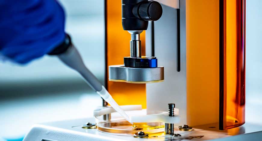
Aresearch team at the University of Nottingham has developed an endoscopic probe enabling practitioners to three-dimensionally (3D) image the stiffness of individual biological cells and complex organisms to discover and treat cancer earlier.
Cancer cells are softer than regular cells in their early stages, which enables them to squeeze through tight gaps and metastasise throughout the body. During this process, cancer cells modify their surrounding environment and create stiff tumours, which insulate them from outside threats.
By measuring the stiffness of individual cells, the university’s optics and photonics group said the device will be able to evaluate microscopic cellular tissue based on abnormal stiffness at the single cell level inside the human body for the first time.
The device, which follows the engineering faculty at Nottingham University’s development of an ultrasonic imaging system for 3D visualisation of cell abnormalities in 2021, achieves high imaging resolution to detect the stiffness of objects down to billionths of a metre (nanometres) through a physical phenomenon known as Brillouin scattering, in which a laser beam interacts with the natural stiffness of a specimen.
To demonstrate this capability, the team visualised the 3D stiffness of Caenorhabditis elegans (C. elegans), a microscopic organism known scientifically as a nematode. Medical Device Network


