


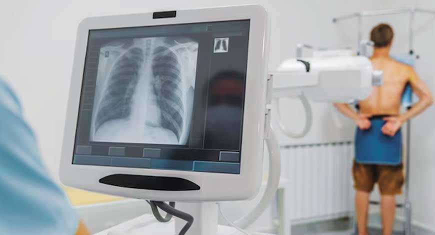
X-ray Equipment
Pioneering the future – Medical imaging solutions unleashed through digital radiography systems
Digital radiography is not just a move but a pioneering leap toward the future of medical imaging solutions, reshaping the landscape of healthcare diagnostics.

The trajectory of medical imaging has undergone a profound transformation with the introduction of digital radiography technology. From the initial discovery of X-rays in the late 19th century to the sophisticated digital imaging systems of today, the evolution has been nothing short of revolutionary.
Replacing conventional film-based imaging, computed radiography employed a phosphor plate to capture X-ray energy. The plates, processed through a laser scanner, produced digital images that can be manipulated for enhanced visualization. While computed radiography marked a significant advancement, its reliance on physical media for image capture posed limitations on acquisition speed and system flexibility.
Digital radiography stands out as a more advanced alternative to computed radiography. Unlike computed radiography, digital radiography eliminates the need for cassettes and chemical processing steps. With digital radiography the radiographic image is captured directly, resulting in faster image acquisition and immediate availability for analysis. This not only enhances workflow efficiency but also reduces radiation exposure. The streamlined process, coupled with the superior image quality inherent in direct digital capture, positions digital radiography as a more advanced and technologically sophisticated solution in comparison to computed radiography.
Traversing the corridors of digital radiography’s evolution reveals the increasing significance of this technology in medical imaging. It serves as a guiding beacon, enabling clinicians to unravel intricate details with unparalleled clarity. The journey of digital radiography, from its nascent beginnings to the present, reflects an unwavering commitment to excellence in diagnostic capabilities.
Innovations in AI-based diagnosis
In the ever-evolving landscape of medical diagnostics, artificial intelligence (AI) is emerging as a transformative force, providing innovative solutions that hold immense potential for revolutionizing the way diseases are diagnosed. Recent developments highlight the ground-breaking strides being made in AI-based diagnostic applications, particularly in chest radiography and the diagnosis of valvular heart disease.
One noteworthy advancement, involves the development of an AI-based model that suggests chest radiography as a potential biomarker of aging. The integration of AI into the analysis of chest radiographs opens up new possibilities for identifying age-related changes and providing valuable insights into the aging process. This not only represents a novel approach to age-related biomarkers but also underscores the versatility of AI in extracting meaningful information from medical imaging.
A further leap in the AI-driven diagnostics is the introduction of a new AI system, poised to revolutionize the diagnosis of valvular heart disease. Traditionally, diagnosing heart conditions, especially those affecting the valves, has been a complex task. The integration of AI introduces a transformative element, potentially streamlining and enhancing the accuracy of valvular heart disease diagnoses. The AI system’s ability to analyze intricate details in medical images, coupled with its capacity to learn and adapt, holds promise for more efficient and precise cardiac evaluations.
Beyond the confines of conventional health records, another breakthrough comes. Here, the focus extends to the application of deep learning in disease diagnosis through image-based phenotyping. This approach represents a paradigm shift, emphasizing the power of AI in extracting phenotypic information directly from medical images. By harnessing deep learning algorithms, this innovative method enhances disease diagnosis by providing a more comprehensive and nuanced understanding of pathological conditions.
These developments collectively underscore the growing influence of AI in reshaping the diagnostic landscape. The integration of AI models not only augments the capabilities of healthcare professionals but also introduces novel approaches to disease identification and monitoring.
In the realm of chest radiography, the AI-based model for aging biomarkers holds significant implications for preventive medicine and age-related healthcare. The chest, being a central hub for vital organs, becomes a canvas for AI to detect subtle changes indicative of the aging process. This innovation opens avenues for proactive health management, allowing for early interventions and personalized care, based on the identified biomarkers.
The transformative impact of AI is perhaps most pronounced in the domain of cardiovascular health. The new AI system, designed for valvular heart disease diagnosis, marks a departure from traditional diagnostic approaches. With the ability to analyze cardiac images with a level of precision and efficiency unparalleled by human counterparts, this AI system not only expedites the diagnostic process but also contributes to more accurate assessments of heart conditions. Such advancements are particularly crucial in the context of cardiovascular diseases, where timely and accurate diagnoses can significantly influence patient outcomes.
Exploring digital radiography technology
Digital radiography has emerged as a transformative force in the realm of medical imaging, revolutionizing traditional radiographic practices. It represents a paradigm shift by allowing for the direct acquisition of X-ray images onto a digital detector. This not only streamlines the imaging process but also enhances the overall quality of diagnostic images. The real-time capabilities of digital radiography offer radiologists a dynamic and accurate tool for visualizing anatomical structures, marking a significant departure from traditional methods.
Digital radiography’s evolution is traced, showcasing its journey from early developments to the sophisticated systems in use today. Beyond its applications in medical diagnostics, digital radiography finds resonance in the realm of material inspection and flaw detection. There is a significance of digital radiography in non-destructive testing (NDT), where its ability to capture detailed images becomes instrumental in assessing the structural integrity of materials across various industries. This cross-disciplinary application underscores the versatility and precision of digital radiography technology.
As we traverse the landscape of digital radiography, it becomes evident that the technology transcends traditional boundaries. Its impact extends beyond medical imaging, permeating industries where precision and reliability are paramount. From real-time imaging advancements in medical diagnostics to the critical role played in ensuring the safety of industrial materials, digital radiography stands as a testament to innovation in imaging technology.
Global market
The global digital radiography market size is estimated at USD 7.38 billion in 2023, projected to hit USD 11.34 billion by 2028, at a CAGR of 9.0 percent during 2023–2028.
Innovative technological advancements and growing awareness of their benefits have led to a measurable, meaningful preference for digital radiography systems. AI-aided X-ray interpretation, now possible due to advancements in computer vision, machine learning (ML), AI, and deep learning algorithms can take the credit for growth. Also, the factors include an aging population, consistently increasing burden of chronic diseases, and the fast-paced adoption of teleradiology in the wake of workflow and staffing challenges are also propelling the market.
Revolutionizing healthcare – The impact of advanced remote-controlled DRF systems RS Kanwar
Managing Director,
Allengers Medical Systems Ltd.
In the dynamic realm of medical technology, advanced remote-controlled digital radiography and fluoroscopy (DRF) systems are ushering in a new era of transformative healthcare.
A standout feature of this equipment is its remote-controlled functionality, a game-changer that prioritizes safety. Medical professionals can operate the equipment from a distance, mitigating radiation exposure for both doctors and healthcare workers. This not only ensures a secure environment but also provides unprecedented flexibility in positioning the imaging equipment, thereby facilitating precise and comprehensive diagnostics for enhanced patient comfort.
Motorized auto-track movements make this equipment swift and easy to position. Exam-specific movement reduces positioning time and makes it very comfortable to use. Advanced image processing techniques with large format flat panel detectors (FPD) produce very high resolution and contrast images. Dynamic FPD gives excellent quality real-time fluoroscopy images that takes this X-ray system to a new level of imaging. A very compact and lightweight design makes this equipment fit well in small rooms without making any compromise on its usability.
Leveraging digital technology for high-resolution image capture, these systems offer unparalleled clarity and detail at lower radiation dose. Large format flat panel detectors cover more patient anatomy, so the procedures become short and lots of radiation is saved.
Efficiency gains from advanced DRF systems are particularly noteworthy. Rapid image acquisition and processing significantly reduce patient waiting times, enabling healthcare providers to deliver prompt and effective care. Features like real-time image stitching and dual energy X-ray enhance efficiency, providing a more comprehensive understanding of anatomical structures and abnormalities.
In summation, the adoption of advanced remote-controlled DRF systems marks a paradigm shift in diagnostic imaging. From heightened safety and image quality to increased efficiency and sustainability, these systems are pivotal in advancing healthcare delivery. As technology progresses, these innovations promise a future where diagnostic imaging is not only more accurate but also more accessible and patient-centric, aligning seamlessly with the evolving needs of the healthcare landscape.
In addition, the increase in digitization in the healthcare sector has led to the increase in the demand for digital radiography market across the globe.
The major restraint of the market is the high costs of digital radiography systems. Additionally, concerns around radiation doses and infrastructure act as challenging factor responsible for stalling growth of the market. Another major restraining factor is the lack of skilled personnel in remote or rural healthcare settings.
Regional insights. North America dominated the overall industry and accounted for the largest share of 40.2 percent in 2022. The rise in the number of patients with orthopedic and cancer diseases is a reason for the region holding the largest chunk of the market share. Additionally, technological advancements in the devices, and a large number of key players with new product development and expansion, are propelling this market.
The Asia-Pacific region is the fastest-growing region in the digital radiography market. China, India, and Japan hold the highest market share in Asia-Pacific. The increasing prevalence of breast cancer, increasing medical costs, an increase in the number of breast cancer awareness initiatives, and significant government expenditure on breast cancer research studies in various Asia-Pacific nations are all boosting the market.
The digital mobile X-ray devices market is estimated at USD 3750 million in 2023. The worldwide adoption of digital mobile X-ray equipment is seeing a significant boom, driven by technical improvements and an increasing prevalence of chronic illnesses.
Advanced imaging technologies
Digital radiography has undergone a transformative evolution, propelled by advanced imaging technologies that enhance diagnostic capabilities. Central to this progression is the integration of flat-panel detectors, a key component revolutionizing the way X-ray images are captured and processed.
Flat-panel detectors in modern digital radiography, unlike traditional image receptors, directly convert X-rays into electronic signals, bypassing the need for intermediate steps. This direct conversion results in improved image quality, enhanced sensitivity, and a more efficient workflow.
Delving deeper into the intricacies of digital radiography, how radiology works compares direct and indirect digital radiography. The direct approach involves the use of flat-panel detectors, capturing X-ray photons and converting them into electrical signals without the need for additional components. In contrast, the indirect method utilizes scintillators to convert X-rays into light, which is then transformed into electronic signals. The direct approach, with flat-panel detectors at its core, emerges as a more streamlined and technologically advanced solution, offering superior image quality and efficiency.
Innovation goes beyond conventional boundaries
New remote-controlled RF tables emerged as a platform for cutting-edge revelations. This unveiling marked a significant stride in enhancing radiographic procedures, offering remote control capabilities that streamline workflow and bolster efficiency. The integration of remote control not only ensures precision in positioning but also aligns with the evolving demands for seamless and user-friendly radiographic equipment.
Complementing these advancements, the next generation of digital mobile X-ray systems, reshape the landscape of point-of-care imaging. This innovation goes beyond conventional boundaries, redefining workflow paradigms. It highlights the system’s capability to offer unprecedented flexibility and agility, making it a game-changer in providing on-the-spot imaging solutions. Such mobility aligns with the contemporary emphasis on accessibility, especially in critical and time-sensitive healthcare scenarios.
Advanced capabilities of DEXA
Dual-energy X-ray absorptiometry (DEXA) has emerged as a cornerstone, offering profound insights into diverse medical applications. The focus is on the versatile utility of DEXA in the clinical evaluation of Cushing’s syndrome. This diagnostic tool plays a pivotal role in assessing and monitoring central obesity, providing clinicians with indices for evaluating changes in adipose tissue distribution during different phases of the syndrome. BMD and visceral fat area (VFA) analyses are highlighted as clinically relevant parameters, although caution is advised regarding potential precision errors in lumbar spine BMD estimation.
DEXA operates by emitting X-rays at two energy levels, allowing for precise differentiation between bone and soft tissue components. It emphasizes the superior accuracy of DEXA in measuring bone density, compared to traditional methods, making it a preferred choice for assessing conditions like osteoporosis and conducting body composition analyses.
Simultaneously, the focus shifts to the evaluation and innovations in digital radiography for non-destructive testing (NDT) purposes. The application of digital radiography in industrial contexts, aiming to detect and characterize material defects without causing damage, and the importance of advanced imaging technologies in NDT, providing a comprehensive overview of how digital radiography, contributes to the field.
Way forward
As we look to the future, the trajectory of digital radiography continues to evolve, promising even greater advancements in medical imaging and diagnostic capabilities. Its emergence opens new frontiers in real-time insights, providing a dynamic perspective for enhanced diagnostics. As technologies like AI, DDR, and advanced detectors continue to mature, we anticipate a future where medical imaging not only meets but exceeds the evolving demands of precision, efficiency, and accessibility in healthcare diagnostics.


