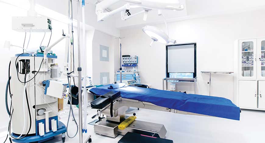
X-ray Equipment
In the Indian context, CR is here to stay

Given the low initial investment, compatibility with a wide range of traditional systems, and effectiveness for smaller clinics, CR has relevance, albeit a niche one.
X-rays were discovered in the late 1800s and shortly after, physicians began using them in medical procedures to identify the presence of metal in the human body. Nobel Prize winner, Marie Curie, really pushed this into the spotlight when she began using her x-ray methods to help doctors in or near battlefields to identify shrapnel and bullet fragments in the injured soldiers. X-rays remained primarily in the medical field for decades.
The story of computed radiograph (CR) in the realm of imaging technology is one of continuous innovation and ground-breaking advancements. With the advent of modern imaging technology, CR has revolutionized the field by providing enhanced and more efficient image capture, processing, and analysis. One of the key revolutions in CR technology is the shift from traditional film-based imaging to digital imaging. This transition has allowed for greater flexibility and improved image quality, ultimately leading to more accurate diagnoses and treatments in the medical field.
Furthermore, CR has also played a significant role in reducing the environmental impact associated with traditional film-based imaging. By eliminating the need for chemical processing and darkrooms, CR has emerged as a more sustainable and environmentally-friendly alternative. Within the realm of computed radiography, another revolution in imaging technology has been the development of wireless digital detectors. These detectors have eliminated the need for cables and physical connection to imaging devices, allowing for CR-eased mobility and flexibility in capturing radiographic images.
Basically, CR can be considered as the digital replacement of conventional x-ray film. Imaging plates are used with the same radiographic inspection methods and techniques as film, and are also available in different system classes (image quality), which have different required exposure times. However, with CR it is not just the imaging plate type that affects image quality, the scan settings used by the scanner are also crucial. In particular, the resolution capability of the scanner (basic spatial resolution or SRb) plays an important part in determining image quality.
In computed radiography, when imaging plates are exposed to x-rays or gamma rays, the energy of the incoming radiation is stored in a special phosphor layer. A specialized machine known as a scanner is then used to read out the latent image from the plate by stimulating it with a very finely focused laser beam. When stimulated, the plate emits blue light with intensity proportional to the amount of radiation received during the exposure. The light is then detected by a highly sensitive analog device known as a photomultiplier (PMT) and converted to a digital signal using an analog-to-digital converter (ADC). The generated digital x-ray image can then be viewed on a computer monitor and evaluated. After an imaging plate is read, it is erased by a high-intensity light source and can immediately be re-used – imaging plates can typically be used up to 1000 times or more depending on the application.
New needle-crystalline detector for x-ray CR technology has been introduced in commercial CR systems with different types of phosphor screens. Discovery of CsBr:Eu2+ as an alternative storage phosphor material is described as excellent because it combines high x-ray absorption capability with the potential for needle-like crystal growth. This offers higher sharpness and sensitivity, particularly for 150m thick screens.
Additionally, CsBr:Eu2+ requires lower stimulation energy and can be erased with less power, as compared to others. CsBr:Eu2+ as a storage phosphor is significant because it performs better than the traditional standard (BaFBr1-xIx:Eu2+). It hints at the possibility of significantly improved image quality and signal-to-noise ratio (SNR) in CR systems when using CsBr:Eu2+ needle screens. This improvement could apply to both existing CR readers and new generations of CR readers.
Fujifilm succeeded in digitizing x-ray photography for the first time in the world, and named the system FCR (Fuji Computed Radiography). The company provided the systems, which can generate high-resolution x-ray images. Having pioneered the world’s first digital x-ray system in 1983, Fujifilm has maintained its focus on building technological innovations, while continuously offering new solutions to the evolving medical industry.
Since the shift away from screen-film radiography, imaging facilities have two basic choices for digital radiography systems – CR or DR. With ongoing technological advancements and the significant reduction in price, DR is rapidly becoming the preferred choice.
CR cassettes use photo-stimulated luminescence screens to capture the x-ray image, instead of traditional x-ray film. The CR cassette goes into a reader to convert the data into a digital image. Digital radiography (DR) systems use active matrix flat panels consisting of a detection layer deposited over an active matrix array of thin film transistors and photodiodes. With DR, the image is converted to digital data in real time and is available for review within seconds.
While both CR and DR have a wider dose range and can be post-processed to eliminate mistakes and avoid repeat examinations, DR has some significant advantages over CR. DR improves workflow by producing higher-quality images instantaneously, while providing two to three times more dose efficiency than CR.
The good and bad of CR is that it enables digital imaging with the traditional workflow of x-ray film. With CR, like film, no synchronization to the generator is required, which had been a requirement for DR imaging. However, recent advances in DR panels are improving their flexibility, portability, and affordability.
DR offers superior throughput compared to CR because it embeds the imaging-processing cycle in the acquisition task – images can appear as quickly as five seconds. CR involves more steps because cassette processing takes longer. Consequently, DR improves workflow because more images can be taken and processed in the same amount of time, allowing imaging facilities to handle more patients in a given period of time.
Both GOS (gadolinium oxysulfide)- and CsI (cesium iodide )-based DR detectors have higher dose efficiency than CR. When DR with CsI is used, DR systems are two to three times more efficient at converting dose to signal than CR. This in CR eased dose utilization means that DR can produce the same image quality as CR at a lower dose or that DR can produce higher contrast resolution images than CR using the same dose.
CR uses a cassette-based system like analog film and is more commonly considered to be a bridge between classical radiography and the increasingly popular fully digital methods.
Considered the most cost-friendly choice, CR is used almost exactly like conventional film, and so requires few changes to the workflow, and requires a smaller initial investment compared to DR devices of similar quality. CR systems also do not speed up your workflow in the same way as a DR panel, and can require more maintenance. CR cassettes may additionally run the risk of getting damaged, but are much cheaper to replace.
In the Indian context, CR’s low initial investment, its compatibility with a wide range of traditional systems, its effectiveness for smaller or low-volume clinics, and multiple sizes allowing for greater flexibility gives it an advantage.
The global market for CR systems is on a trajectory of steady growth. Based on the recent study by Growth Plus Reports, the global CR market is projected to reach USD 6.45 billion by 2031 from USD 4.40 billion in 2022, at a CAGR of 4.9 percent.
In the market adoption of this, CR technology is also fuelled due to the growing emphasis on lowering radiation exposure during medical imaging operations. CR technology frequently used in medical imaging produces high-resolution images of soft tissues, bones, and organs and, as compared to conventional film-based radiography, it offers significant advantages including the capacity to electronically store and transfer digital images, quicker image capture times, and reduced radiation doses for patients.
According to the WHO, over 1.71 billion individuals worldwide suffer musculoskeletal problems, and rise in orthopedic disorders and chronic diseases worldwide is boosting the demand for computed radiography. Some other factors, driving the revenue growth of the CR market are the development of high-resolution imaging systems, improved image analysis and processing software, and the integration of artificial intelligence (AI) and machine learning algorithms to advance diagnostic accuracy.
In addition, the market revenue for CR is growing due to the development of portable and wireless devices, and improvements in CR hardware and software. Portable CR systems are
gaining popularity due to their ease of use, portability, and cost-effectiveness. These systems are particularly useful in remote and rural areas where access to medical imaging services is limited.
The orthopedic segment is having the highest growth rate due to the factors, such as fractures, osteoporosis, and arthritis, particularly in elder people. For orthopedic applications, like bone fractures and joint replacements, CR is a cost-effective imaging technique. In addition, orthopedic diagnosis highly demand CR technology due to improved image quality, reduced radiation exposure, and in CR eased workflow efficiency.
The cancer diagnostic segment is poised for significant revenue growth due to the global rise in cancer cases. Computed radiography, with its improved features, has become vital in early cancer detection, helping radiologists identify tumors accurately and quickly. Compared to MRI and CT scans, CR offers a cost-effective imaging solution, driving its demand in cancer diagnosis. The hospitals segment and diagnostic centers are also demanding CR systems.
Evolving dynamics in the Indian computed radiography systems industry
 Chandershekhar Sibal
Senior Vice-President & HOD of Medical Division,
FUJIFILM India
Step into the future of medical imaging with computed radiography (CR) systems. Utilizing cassette-based phosphor storage plates, this technology transforms images into a digital format, making them readily available for processing, archiving, and presentation. According to a report, the digital X-ray market in India is forecasted to grow from USD 375.40 million in 2023 to USD 610.65 million by 2028. This highlights the substantial progress occurring in the Indian computed radiography systems sector, spearheaded by FUJIFILM India’s technical innovations.
The Indian healthcare industry has been expanding, driving the demand for medical imaging solutions like CR system. CR serves as the primary entry point for the adoption of digital imaging technology in many imaging centers, thanks to its economical setup. It seamlessly integrates with most existing conventional systems and can be easily retrofitted to pre-existing installations. Many computed radiography systems, specifically FUJIFILM India’s computed radiography (FCR), offer versatility by accommodating cassettes of various sizes, allowing for detector selection tailored to the specific procedure, and enhancing positioning flexibility across examination areas. Furthermore, compact single-plate readers efficiently manage high patient volumes, ensuring a continuous workflow where one image is processed while the next is acquired in rapid succession. Similarly, computed radiography requires fewer alterations in workflow, potentially leading to cost savings. This is useful for converting analog fixed X-ray machines, analog mobile X-ray, and analog mammography machine output to digital.
Challenging the conventional computed radiography technology
For this market segment, FUJIFILM India presents an ideal solution with their cutting-edge FUJI computed radiography (FCR) line, featuring products like the Prima T and Prima Tm. These small, tabletop reader units boast high-speed processing, dismissing the common misconception of slow processing in CR. They showcase a compact, space-saving design, offer swift processing rates of up to 57 IPS per hour (or up to 73 IPS per hour for the Tm model), and provide various cassette size choices. The Tm model, in particular, has gained recognition as the world’s most compact all-in-one tabletop CR, featuring standard mammography image reading capabilities.
Overall, the introduction of innovative CR systems, including wireless, portable, and image-processing software, has boosted CR demand. These systems offer better image quality, patient comfort, and faster processing. Additionally, a robust healthcare infrastructure and key players contribute to regional revenue growth.
However, the installation and maintenance of CR systems require a significant upfront investment, restricting the revenue growth of the CR market. CR involves exposure to ionizing radiation, which can pose health risks to patients and healthcare workers, hampering the revenue growth of the global CR market.
The major players operating in the global CR market are Siemens Healthcare, Konica Minolta, Inc., Jones x-ray, Inc., GE Healthcare, Agfa-Gevaert Group, Koninklijke Philips N.V., FUJIFILM Corporation, Imaging Dynamics Company Ltd., Detection Technology Plc., Desert Health Imaging Technologies., and Strategic Developments. The players are launching new technologically advanced products into the market.

Artificial Intelligence in computed radiography
CR has undoubtedly undergone transformative advancements in imaging technology, leading to a revolution in the field. The integration of AI algorithms in CR systems has significantly enhanced the ability to detect and highlight abnormalities in images, assisting radiologists in making accurate diagnoses and improving patient outcomes. Aside from the overall improvements in CR technology, another revolutionary advancement that has emerged is the integration of AI algorithms that can automatically detect and highlight abnormalities in images, allowing radiologists to focus their expertise on interpreting findings rather than spending excessive time searching for potential issues. This not only accelerates the diagnostic process but also enhances the accuracy of diagnoses.
Despite all challenges, CR continues to revolutionize radiology, enhancing patient care and outcomes. With emerging trends and future developments, such as digital radiography and point-of-care solutions, the future of medical imaging looks promising, underlining its pivotal role in modern healthcare.
CR and PACS (picture archiving and communication systems) are gaining popularity among medical professionals, promising economic benefits and increased productivity. Recent technological advancements have further improved image clarity and processing speed, expanding its clinical applications.
However, as with any imaging technology, artifacts can compromise the accuracy of radiological diagnosis. Artifacts in CR may differ from those in traditional radiography, and they can arise from various components of the system, including displays, imaging plates, plate readers, image processing software, and operator errors.
Some common types of artifacts in CR system include lead oxide transmission from lead screens, atmospheric dust/dirt (ADD), damage to lead screens, fingerprints, scratches on imaging plates, electromagnetic interference, and inadequate erasure. These artifacts can affect image quality and interpretation.
Atmospheric conditions, such as air dust and debris, can introduce ADD artifacts. ADD can cause the imaging plate to stick, resulting in white artifacts on grayscale displays. Lead screens, when used without protective coatings, can transfer lead oxide to the imaging plates, requiring daily cleaning.
Electromagnetic interference, though less common, can create alternate row intensity artifacts in CR images. These artifacts occur when some significant interference, such as a power line, is nearby. Shielding or relocating equipment can reduce their impact.
Bent imaging plates are more likely to break and can introduce artifacts. Backscatter from objects behind cassettes can also create artifacts. Lead foil on the cassette’s back may help mitigate this issue.
Plate reader artifacts can arise if imaging plates are not properly erased before reuse. The erasing cycle for exposures with mistakes should be longer and more powerful. Image processing is crucial for producing high-quality images in CR. Inappropriate processing parameters can introduce artifacts, and operator errors in selecting settings can affect image quality.
The light bulb effect occurs when radiation from the patient’s bed penetrates the imaging plate. This artifact is more pronounced in obese patients or when x-ray beams are not properly collimated. Proper kilovoltage and collimation can reduce its occurrence.
Double-exposure artifacts can result from radiographers taking consecutive exposures. This error can lead to incorrect diagnosis and legal consequences. CR offers the advantage of reducing radiation dose without compromising diagnostic accuracy. However, foreign body artifacts, such as clothing or adhesive tape, can sometimes be mistaken for pathology.
Overall, while CR and PACS offer numerous benefits, understanding and addressing artifacts are essential for accurate diagnosis. Artifacts can arise from various sources within the system and can significantly impact image quality and interpretation. Proper equipment maintenance, quality control, and operator training are crucial for minimizing artifacts in CR systems.


