
MB Stories
Rethinking MRI to provide the reach that it deserves
Cutting-edge technology expands imaging access, transforming diagnostics for all, where MRI innovations redefine medical standards globally. And yet, the global disparity of MRI is a major challenge, with many LMICs experiencing limited access.

In recent years, significant strides have been made in the field of MRI, particularly with the development of groundbreaking technologies aimed at enhancing accessibility and performance. These advancements have the potential to revolutionize healthcare by providing cutting-edge imaging solutions to previously underserved populations worldwide.
The global disparity of MRI is a major challenge, with many low- and middle-income countries (LMICs) experiencing limited access to MRI. The reasons for limited access are technological, economic, and social. They face severe personnel and imaging equipment shortages. For MRI the gap is considerably more significant. Along with this equipment deficit, a severe labor shortage affects medical physicists, radiologists, and radiographers, with 1.9 versus 97.9 radiologists per million people in low- versus high-income countries, respectively.
In a groundbreaking development, India-based Voxelgrids recently launched an advanced and affordable MRI scanner with the potential to revolutionize healthcare accessibility not only in India but also worldwide. With funding from BIRAC (Biotechnology Industry Research Assistance Council), an institution set up by the Department of Biotechnology (DBT), Government of India, to nurture and promote biotechnology innovations and industry in India, Voxelgrids has successfully developed a cutting-edge MRI (magnetic resonance imaging) scanner that is significantly more affordable than traditional counterparts, breaking down barriers to access for billions of people previously unable to afford the procedure.
In 2021, the company closed on its series-A funding with Zoho Corporation, a privately held multi-billion-dollar software company. Voxelgrids today is an independent entity supported by multiple stakeholders and is on a mission to provide cost effective, state-of-the-art magnetic resonance imaging scanners for the world markets.
The Voxelgrids MRI scanner, priced at approximately USD 400,000, represents a fraction of the cost of conventional 1.5T whole-body scanners, which typically range from USD 670,000 to USD 1.2 million. The substantial cost difference can create a profound impact on healthcare accessibility worldwide.
A multi-institutional project on the development of the 1.5T superconducting MRI scanner (IMRI Project) had been initiated by the Ministry of Electronics & Information Technology (MeitY) through SAMEER-Mumbai, the nodal agency. The partnering institutes are Inter-University Accelerator Centre (IUAC)-New Delhi for 1.5T superconducting magnet and 4K ZBO cryostat, SAMEER-Mumbai for RF coils/amplifiers and other associated RF electronics, spectrometers, couch and the integration of the scanner, CDAC-Kolkata, and CDAC-Trivandrum for the imaging software, and DSI-Bangalore for MR physics.
IMRI team of IUAC has designed every component of the 1.5T superconducting MRI system through an extensive electromagnetic, electrical, thermal, structural, and cryogenic simulation, using various commercial FEA software. The IMRI magnet consists of eight superconducting coils having six primary coils and two active shield coils made of Cu/NbTi conductors having a critical temperature of 9.2K. The superconducting coils are wound onto a metallic bobbin structure having precisely machined winding pockets. The coils are wound onto each winding pocket using a predefined winding tension following an extensive electrical insulation scheme. An efficient quench protection system has been developed to dissipate its entire stored energy onto the entire magnet in a controlled manner through a self-activated electrical circuit. An efficient external interference screening (EIS) system has been designed and developed for shielding the imaging volume from any low-frequency external magnetic disturbances.
A team of researchers from the NYU Langone Radiology – Center for Biomedical Imaging convened over four days to build a functional MRI machine from scratch. The results laid the groundwork for a more equitable model of making medical imaging technology accessible to underserved areas around the globe.
A typical clinical MRI scanner weighs up to 17 tons and can cost as much as USD 3 million. Each machine requires three dedicated rooms – one for scanning, one for electronic attachments, and another to scan the patient. Its size, cost, and the expertise required to operate it put it out of reach of many healthcare providers in the developing countries.
In just four days, the team created an entire MRI machine. To put their model to the test, the team used what is known as a phantom, a simple water-filled test object whose exact dimensions and contents were known only to one member of the team. If the machine produces an accurate image, it indicates that the scanner works properly. After a few false starts, the DIY machine yielded clear images. The team is now making all the designs for the components, many of which were 3D printed, as well as the software developed and tools used for designing the magnet, publicly available on their project website.
Efforts are also underway to enhance the accessibility and affordability of MRI services, particularly in low-resource settings. Initiatives aimed at developing cost-effective imaging solutions and establishing collaborative networks for knowledge sharing and capacity building are crucial steps toward ensuring equitable access to essential healthcare services. By leveraging innovative financing models and strategic partnerships, stakeholders can work together to overcome barriers and expand the reach of MRI technology to underserved communities worldwide.
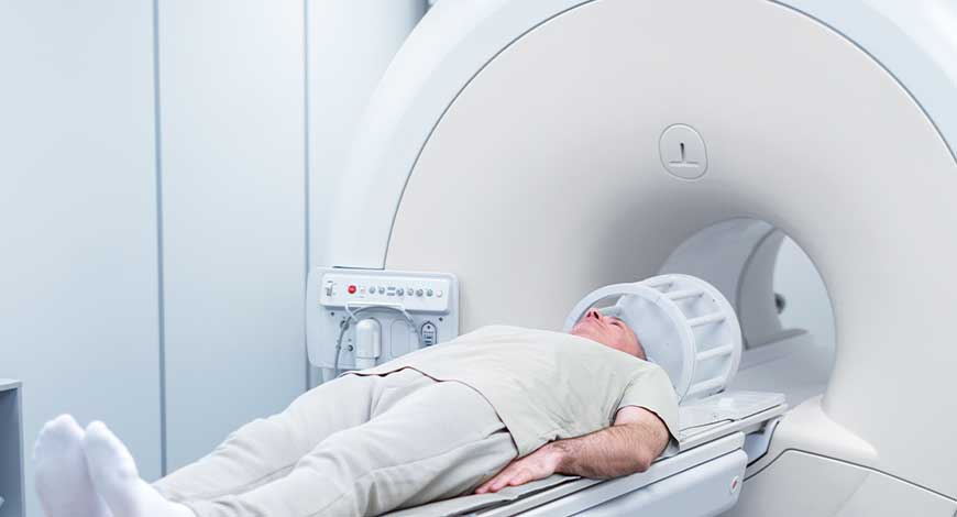
That said, even in the United States, getting an MRI may require days of waiting and a midnight drive to some distant hospital. The patient must come to the scanner, not the other way around. An MRI scanner employs a magnetic field to twirl atomic nuclei in living tissue – specifically the protons at the heart of hydrogen atoms – so that they emit radio waves. To generate the field, a standard scanner employs a large, powerful superconducting electromagnet that pushes a machine’s cost to USD 1.5 million or more, pricing MRI out of reach of 70 percent of the world’s population.
Indian market dynamics
The Indian MRI market in 2023 is estimated at ₹1800 crore. Presently, in India, there are 1½–2 MRI machines per million population whereas in the developed countries it is 10–30 MRIs per million population. High costs, maintenance issues, power consumption, and the requirement for specialized expertise together pose obstacles to the widespread access to MRI services in India, especially in rural and underserved regions.
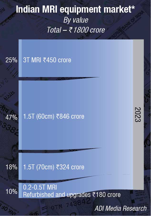
By segment, while 3T systems hold a 25-percent market share, it is the 1.5T systems that are the workhorse. And within the 1.5T category, while the 60-cm systems dominate, it is the 70-cm systems that are preferred over the 60-cm models. The refurbished, the upgrades, and the 0.2T–0.5T categories are shrinking.
The Indian market is expected to grow at a CAGR of 7 percent over the next five years. The anticipated developments in MRI systems in India, including advanced portable and cost-effective MRI solutions, hold the potential to enhance accessibility, especially in remote regions. Furthermore, the integration of AI for image analysis and interpretation stands to elevate diagnostic precision and streamline processes.
|
Leading players-Indian MRI equipment market |
|
| Tier 1 | Siemens, Philips and GE |
| Tier 2 | United Imaging |
| Others | Fuji (Hitachi) & Canon (formerly Toshiba); Phantom (refurbished) |
|
*Vendors are placed in different tiers on the basis of their sales contribution to the overall revenues of the Indian MRI equipment market. ADI Media Research |
|
Global market dynamics
The global MRI equipment market is estimated to reach USD 49.40 billion by 2030, from USD 16.72 billion in 2023, growing at a CAGR of 16.7 percent, according to Coherent Market Insights. This non-invasive test produces high-quality images of the body’s internal structures without using harmful ionizing radiation. Today, MRIs are the preferred method for diagnostic imaging of soft tissue structures like the brain, spinal cord, musculoskeletal system, small bones, liver, and pelvic area. They can also detect abnormalities in vascular structures, such as blood vessels.
Market trends
An important factor that is gaining traction is the focus on development of helium-free MRI systems. Helium is an essential component in MRI systems and the severe shortage of helium worldwide has made the manufacturing of MRI systems expensive. To overcome this, vendors are focusing on helium-free MRI systems. By eliminating the reliance on helium, which is essential for cooling superconducting magnets in conventional MRI systems, these next-generation scanners mitigate concerns related to helium scarcity and supply chain disruptions. This innovation not only enhances operational efficiency but also reduces environmental impact and operating costs, making MRI imaging more sustainable in the long term.
Many market players are constantly engaged on providing open MRI architecture, which are extremely suitable for oversized patients and the patients that suffer from claustrophobia. In order to make these machines attractive for children, and to make the experience fun for children, some of these machines have cartoons or drawings on them. The amount of anxiety is reduced with the use of open systems, and due to which this segment becomes more effective. That said, on the basis of architecture, the closed segment is expected to have a larger market share in the coming years. This segment makes use of magnetic fields that are powerful and offer higher frequency of radio waves. These two factors help in obtaining detailed images. The analysis obtained with the help of this type of architecture offers less chances of errors and it also gives a detailed selection of slice. There are chances that the patients feel disturbed due to the presence of loud noise and they may also feel claustrophobic. These are some of the reasons due to which the results could be inaccurate.
The vision quality is good as compared to use of other types of machines and the faster timing in acquiring images also leads to the growth of the market. Market players across the world are engaged in increasing the number of innovative products introduced in the market.
The applications of high-field MRI systems in brain imaging are a major factor contributing to growth of global MRI systems market. High-field MRI systems provide excellent spatial resolution and signal-to-noise ratio, which allow clinicians to obtain very detailed anatomical and functional images of the brain. Many neurological and psychiatric disorders, such as Alzheimer’s, dementia, multiple sclerosis, epilepsy, autism, and brain tumors are being increasingly studied by using advanced imaging techniques supported by high-field MRI scanners.
Adoption of portable and mobile MRI systems is having a significant influence on the global MRI market. Mobile and portable MRI systems allow for MRI scans to be conducted outside of traditional hospital settings, thus bringing the important diagnostic technology directly to the patient. This represents a paradigm shift in how MRI exams are performed.
Emergence of wide-bore MRI systems is having a significant impact on the market too. Wide-bore MRI systems have a larger opening or bore, which makes them more accessible and comfortable for certain patient groups. The larger opening reduces feelings of confinement, which is more accommodative for claustrophobic or anxious patients. It also enables the scanning of larger body parts that may not fit inside the standard MRI machine, such as obese individuals or those undergoing whole-body imaging.
Refurbished MRI systems, given their low costs continue to sell to a niche market. Small diagnostic centers and standalone hospitals opt for refurbished systems.
Restraints
The exorbitant cost of MRI systems poses a significant challenge for the widespread adoption of MRI technology. MRI systems require huge capital investments, with high-end machines often costing over USD 1 million. Moreover, additional costs are incurred toward site preparation, installation, maintenance, and service contracts over the lifespan of the systems. The operating expenses of MRI scans are also high due to large overhead costs associated with high-field strength magnets and other advanced imaging hardware.
Strict regulatory approval for MRI systems is significantly hampering the growth of the global MRI market. Regulatory agencies across most countries have implemented rigorous guidelines and standards for approving new MRI systems and upgrades to the existing systems before they can be commercially launched. This lengthy approval process has increased the time taken by MRI manufactures to introduce innovative new models in the market.
Technological advances in MRI systems are likely to drive the market
Advancements in MRI hardware and software have led to significant improvements in image quality and acquisition speed. Cutting-edge MRI systems now boast enhanced signal-to-noise ratios, resulting in sharper and clearer images that enable more accurate diagnoses. Furthermore, the development of accelerated imaging techniques has substantially reduced scan times, with some protocols promising comprehensive MRI exams in as little as five minutes. This rapid imaging capability not only enhances patient comfort but also optimizes workflow efficiency.
Innovative coil designs have also played a pivotal role in enhancing MRI performance and versatility. Advanced coil arrays, coupled with sophisticated signal processing algorithms, enable targeted imaging of specific anatomical regions with unprecedented detail and sensitivity. Moreover, flexible coil configurations facilitate imaging of challenging patient populations, such as pediatric and claustrophobic patients, while ensuring optimal image quality and diagnostic accuracy.
Furthermore, the integration of AI into MRI workflows holds immense promise for improving diagnostic accuracy and efficiency. AI algorithms can automate time-consuming image analysis tasks, such as image segmentation and lesion detection, thereby enabling radiologists to focus on interpretation and clinical decision-making. Moreover, AI-based reconstruction techniques enable real-time image enhancement and artifact suppression, further enhancing the diagnostic utility of MRI imaging.
In the long run, advanced technologies as the new MRI system features as innovative workflow solutions, image enhancement, and accelerated scan technology, which together contribute to reducing the time required for MRI procedures, can significantly drive growth.
Advancements in MRI technology extend beyond clinical applications, encompassing areas, such as neuroscience research, drug development, and personalized medicine. By providing researchers with unprecedented insights into anatomical structures and physiological processes, advanced MRI techniques empower scientific discovery and innovation across diverse disciplines.
Some recent innovations
Real-time tracking of moving tumors using MRI during proton therapy. On January 9, 2024, a scientific prototype for MRI-guided proton therapy was inaugurated in Dresden, UK. With this installation, experts from the fields of medicine, medical physics, biology, and engineering are embarking on the scientific testing of a new form of radiotherapy for treating cancer. For the first time globally, a full-body MRI device for real-time imaging is combined with a proton therapy system in the form of a prototype.
Once cancer patients are monitored during their radiation treatment, using real-time MRI imaging, it significantly improves the targeting accuracy of proton therapy. A globally unique combination of a full-body MRI machine that rotates around the patient for real-time imaging and a proton therapy system was created in Dresden. Scientific operation has now begun in January 2024.
MRI’s advantage over conventional imaging modalities is that it can visualize the tumor in higher contrast. This makes it possible to better delineate the tumor from surrounding healthy tissue and to define the volume to be irradiated more accurately. Furthermore, MRI imaging can visualize any potential changes in the shape and size of the volume to be irradiated between consecutive radiation sessions. This enables the beam application to be adjusted individually and immediately. In addition, it allows the real-time MRI imaging to visualize tumor movement during a radiation session and to synchronize it with the radiation application. The prototype that has now been installed will be the first of its kind globally to investigate the extent to which the accuracy of proton therapy can be improved with the help of full-body real-time MRI imaging.
A new MRI technique that is more precise and 25 times faster. MRI scanners can be programmed to measure myelin, which is the insulating layer that protects neurons and is a key indicator of numerous neurological disorders. However, myelin imaging is extremely slow, traditionally requiring the MRI scanner to collect huge amounts of data. The University of British Columbia researchers have developed a new technique that collects a tiny fraction of the data while providing higher-quality myelin measurements.
This new method is called the constrained, adaptive, low-dimensional, intrinsically precise reconstruction (CALIPR) framework. The technique allows for the acquisition of advanced, high-resolution quantitative maps of the human brain in 7.5 minutes, instead of 3 hours, making myelin imaging approximately 25 times faster than a conventional full MRI scan. The new framework has demonstrated excellent precision in quantifying myelin content, with low variability in measurements for both the brain and the spinal cord. Compared to existing techniques, CALIPR is much more efficient and can potentially improve the detection of demyelinating diseases, such as multiple sclerosis.
Magnet upgrade helps fMRI probe neuron clusters. Neuroscientists at UC Berkeley and the BRAIN initiative from the National Institutes of Health have developed a series of technological improvements that dramatically increase the spatial resolution of an fMRI (functional magnetic resonance imaging) machine. The improvements allow an fMRI to image voxels – the 3D equivalent of pixels – that are less than half a millimeter on each side. In doing so, they have passed an important threshold relative to the structure of the human brain, to roughly match the scale of functional clusters of neurons.
The NexGen 7T scanner is a ten-fold improvement in resolution over 7T MRI machines that are currently available to researchers. The finer scale could be useful for many research questions. Any fMRI scanner works by creating magnetic fields that affect the orientation of molecules in the brain. Once one magnetic field is set to a predictable pattern, densities or types of tissue can be differentiated by applying a second magnetic field and rapidly oscillating the orientation. Various techniques have been developed, which use blood flow or oxygen levels as a proxy for brain activity.
The team chose to work with a 7T MRI scanner in part to strike a balance between technical requirements and availability, compared to even more powerful, experimental MRI machines. Stronger magnetic fields can not only increase the potential for high-clarity images, but also increase the potential dangers of RF heating for people inside the scanner. So while only one NexGen scanner has been built so far (at UC Berkeley), any of the 100 or so operational 7T MRI scanners globally could in theory be retrofitted to the same specifications.
Clinical applications with MRI
MRI scanners have become indispensable tools in clinical practice, offering unparalleled insights into the human body’s structure and function through ongoing research and innovation.
One significant application of MRI scanners lies in cardiovascular imaging. With its exceptional soft tissue contrast and ability to visualize blood flow dynamics, MRI plays a crucial role in diagnosing and monitoring cardiovascular diseases. Researchers have developed advanced MRI techniques, such as cine MRI and magnetic resonance angiography (MRA), to assess cardiac function, detect arterial stenosis, and evaluate myocardial perfusion. These techniques provide clinicians with comprehensive information for accurate diagnosis and treatment planning in patients with conditions, such as coronary artery disease, heart failure, and congenital heart defects.
MRI scanners have emerged as valuable tools in neurological imaging, enabling non-invasive assessment of the brain and spinal cord. Advanced MRI sequences, including diffusion-weighted imaging (DWI) and functional MRI (fMRI), offer insights into brain structure, connectivity, and function. Clinicians utilize MRI to diagnose neurological disorders, such as stroke, multiple sclerosis, and brain tumors, guiding treatment decisions and monitoring disease progression over time.
In the realm of oncology, MRI scanners play a crucial role in cancer diagnosis, staging, and treatment planning. Through techniques like dynamic contrast-enhanced MRI (DCE-MRI) and diffusion tensor imaging (DTI), clinicians can characterize tumor morphology, assess vascularity, and evaluate treatment response. MRI-guided biopsy procedures further enhance the accuracy of cancer diagnosis, enabling targeted tissue sampling for histopathological analysis.
MRI scanners offer valuable insights into musculoskeletal disorders, facilitating the diagnosis and management of orthopedic conditions and sports injuries. Advanced MRI techniques, such as magnetic resonance arthrography (MRA) and cartilage-sensitive imaging, enable detailed assessment of joint structures, ligaments, and cartilage integrity. Clinicians utilize MRI to detect ligament tears, cartilage defects, and bone abnormalities, guiding surgical planning and rehabilitation strategies for optimal patient outcomes.
In respiratory medicine, MRI scanners have emerged as powerful tools for imaging the lungs and airways, offering non-invasive evaluation of lung function and pathology. Novel MRI techniques, including hyperpolarized gas imaging and magnetic resonance elastography (MRE), enable assessment of lung ventilation, perfusion, and tissue stiffness. Clinicians utilize MRI to diagnose and monitor respiratory conditions, such as chronic obstructive pulmonary disease (COPD), asthma, and interstitial lung disease, guiding treatment decisions and assessing treatment response over time.
Moreover, MRI scanners play a vital role in pediatric imaging, offering radiation-free imaging options for children with diverse medical conditions. Pediatric MRI protocols are optimized to minimize scan times and ensure patient comfort, making MRI an ideal imaging modality for pediatric patients with congenital anomalies, neurological disorders, and musculoskeletal injuries.
Research in MRI technology extends beyond clinical applications to include fundamental studies exploring the biophysical principles underlying magnetic resonance phenomena. These studies provide valuable insights into the interactions between magnetic fields and biological tissues, laying the foundation for the development of novel imaging techniques and diagnostic tools.
Interdisciplinary collaborations between researchers in fields, such as engineering, physics, and medicine are driving innovation in MRI technology. By combining expertise from diverse disciplines, researchers can overcome technical challenges, explore new avenues for research, and translate scientific discoveries into clinical applications that benefit patients worldwide.
The global impact of MRI advancements extends beyond healthcare, influencing various sectors, such as education, industry, and policymaking. As MRI technology becomes more accessible and versatile, its applications transcend traditional boundaries, driving innovation and progress across diverse domains. From improving educational resources for medical students to enhancing industrial quality control processes, the widespread adoption of MRI technology heralds a new era of possibilities and opportunities.
Safety measures and enhancements in MRI technology
The safety of patients and healthcare professionals during MRI procedures is paramount, and advancements in technology continue to address safety concerns and improve overall safety measures. However, when challenges arise they also present opportunities for improvement and innovation in MRI safety measures. Regulatory updates, such as the FDA’s recent revisions to MRI-related guidance documents, provide an opportunity for healthcare facilities to reassess their safety protocols and implement enhanced measures.
Technological advancements offer another avenue for addressing safety challenges. Automated patient monitoring systems, motion detection algorithms, and fail-safe mechanisms integrated into MRI scanners can help mitigate risks and enhance safety during imaging procedures. Advancements in imaging techniques and software algorithms enable clinicians to accurately assess the compatibility of implants with MRI scanners, reducing the likelihood of adverse events.
Outlook
Everyone deserves access to MRI. As an extremely powerful imaging tool, MRI has proven to play a pivotal role in the fight against many of the world’s most prevalent diseases. But the cost and complexity of deploying conventional MRI scanners limits its reach. As a result, more than 50 percent of people on our planet still have no access to this potentially life-changing technology. And those who do have access must often endure long waiting times and travels based on the current availability. So, if we really want to change this, we need to rethink MRI.
Second Opinion:-


