
X-ray Equipment
Technology revolutionizing imaging techniques

It is now possible to create large, flexible X-ray detectors powered using only a couple of coin-cell batteries. Alternatively, one could create portable X-ray detectors that, coupled with portable X-ray sources, could fit into ambulances and be used to diagnose on the road and not just in hospitals.
Radiology technology has advanced by leaps and bounds to a stage where the vendors too understand the impact of the developments toward patient advantage and, therefore, cost-effective solutions are being offered. This will go a long way in helping to promote technology and provide its benefits to Tier-II and Tier-III townships of countries like ours, and other developing countries.

The Indian X-ray equipment market is growing exponentially with an increase in the number of imaging centers. In addition, demand is also expected to come from district hospitals, as the health ministry has proposed to convert 75 district hospitals into medical colleges in the third phase of a scheme, which aims at boosting availability of human resource for the health sector. The upgradation of each of these district hospitals into medical colleges is estimated to cost around ₹325 crore.
The Indian X-ray equipment players comprises a mix of large multinational companies and local companies, and there is a significant overlap of the target segments of both. With the MNCs also bringing in the de-featured products to be cost-competitive, the differentiation tends to largely focus on highlighting the lower cost of ownership, service promptness of the field force, and robustness of the equipment, which is typically seen in small-value products. In the large-value deals, the proposition changes a bit to include other add-ons, such as education and training of the staff on the usage of the equipment, workshops, loan and EMI facilities, pay-per-use model, lease model, and turnkey projects.
With all this focus on value-products, the larger companies still package the innovations, which are out of reach of the local companies, in the premium products offered at premium prices. The hospitals, which strive to position themselves as state-of-the-art hospitals, sporting the latest high-end equipment, are the target customers for these models. The decision-making process that was earlier based on the doctor’s feedback, now also includes other stakeholders, such as the finance, administration, and the biomedical teams. It is changing in tandem with external forces shaping the healthcare-delivery industry. Companies offering performance products at competitive prices are poised to gain significantly from this change.
Indian market dynamics
The Indian market in 2018 is estimated at 13,000 units, with value estimated as ₹860 crore. The CR machines dominate with a 40 percent share, DR contributes 34 percent and analog 26 percent, by value. As expected, by quantity, the analog systems have a 50 percent share, CR constitutes 44 percent, and DR 6 percent.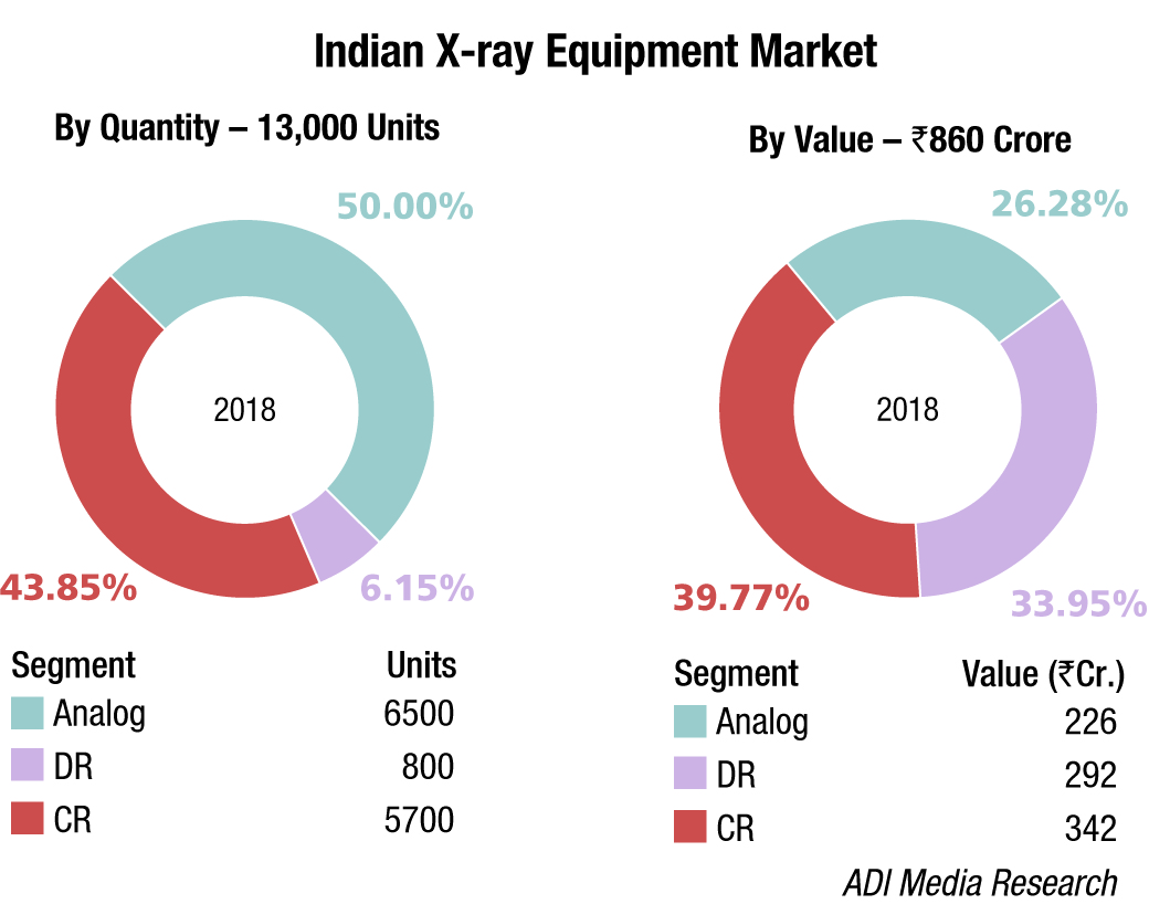
In the analog and DR segments, the competition for market share on a national level is among four brands – Philips, Allengers, GE, and Siemens, albeit Siemens has as a matter of strategy reduced its presence in the mid- and entry-level segment. Skanray and Epsilon did well in 2018. In the CR segment, race is between four brands – Fuji, Agfa, Carestream, and Konica.
The government hospitals, including the ESI and railway hospitals, is replacing its machines from analog to DR. The CR is expected to lose market share, as the price differential between CR and DR is narrowing. 
A handful of discerning customers opt for the ceiling-suspended models, with automatic movement, average unit price being ₹1.5 crore.
Technology trends
Medical imaging saves millions of lives each year, helping doctors to detect and diagnose a wide range of diseases. Because non-invasive early disease detection saves so many lives, scientific investment continues to increase. Let us take a look at the biggest up-and-coming medical imaging technology trends:
| Segment | Tier I | Tier II | Others |
|---|---|---|---|
| Analog | Philips and Allengers | Siemens and GE | Skanray, Epsilon, Kiran Medical (Trivitron), and BPL |
| DR | Philips | GE, Siemens, and Allengers | Konica, Fuji, and Canon (Prognosys) |
| CR | Fuji | Agfa and Carestream | Konica |
| *Vendors are placed in different tiers on the basis of their sales contribution to the overall revenues of the Indian X-ray Equipment market. | |||
| ADI Media Research | |||
Artificial intelligence (AI). AI has the potential to revolutionize the medical-imaging industry by sifting through mountains of scans quickly, and offering providers and patients with life-changing insights into a variety of diseases. Big-name companies are partnering together to explore how AI can improve diagnostics. AI and radiologists will continue to complement each other in future. Interestingly, although AI is faster on its own, the best results come from teamwork – humans utilizing the AI algorithm as a second look. AI will support radiologists and medical imaging departments by streamlining workflows and improving productivity.
Wearables. Wearable medical devices will make more inroads into diagnostic imaging in future. Most recent invention for medical imaging is the MEG wearable brain scanner. It is lightweight and worn like a helmet, and can measure brain activity while people make natural movements, such as drinking, nodding, stretching, and the like. This brings improved imaging possibilities to patients with disorders that cause body movements like epilepsy. Another important wearable device is an MRI glove, worn next to the skin; it can provide clear, constant images of moving joints and tendons without motion artifacts. The benefits of the images produced by the MRI glove include providing a clear map of the anatomy of the hand, aiding in everything from surgery to the design of more accurate prosthetics.
3D imaging and virtual reality. Virtual reality and 3D imaging technologies are not only great for entertainment, but they also have important implications within the medical-imaging industry. MRIs and CT scans have, currently, their display in 2D which requires physicians to use their imagination to mentally stitch together a full picture of the 3D organ or body part. Now, new technologies like Echo pixel true 3D have made it possible for radiologists or physicians to take slices of MRI pictures to create a 3D image that physicians can examine with 3D glasses, a VR headset, or even print using a 3D printer and special plastic.
Outlook
X-rays could be about to change. Since its discovery at the end of the 19th century, the radiation has provided a window into the inner workings of the body, and later gave the power to see inside everything from buildings to suitcases. However, the technology has remained in principle the same. But X-rays could be much more flexible in future. New technology means that X-ray detectors could one day effectively be printed onto any suitable surface. So, they could be made curved to wrap around the part of the patient’s body being scanned to produce a much more accurate image. They could be large enough to scan an entire truck in one go. And they could be portable enough to quickly carry through a crowd to inspect a suspicious package. And this future is now a step closer, thanks to recent advances.
Today, many X-ray images are created by firing the rays onto a digital detector rather than photographic film. These detectors convert the X-rays into electrical charges and a computer converts these into a digital image. Many industries have benefitted from this massive leap from X-ray film because it has enabled the creation of more detailed images that can be viewed in real time. But the solid detectors are also rigid and flat and so limited.
By contrast, the human body naturally consists of curved surfaces. And firing X-rays through a curved object onto a flat detector can cause errors. This can lead to a cancer misdiagnosis or a patient being given the wrong dose of radiotherapy. Overexposing someone to X-rays can also cause tissue damage and even the growth of secondary tumors. Because current X-ray detectors are mostly made from thick slabs of inorganic material, they are expensive to make in large sizes for scanning large objects, such as vehicles. Some detectors also require very high voltages to operate and so would not be suitable for portable applications.
For inspecting manufactured parts and products for defects (known as non-destructive testing), traditional X-ray films are still commonly used to capture images because they are cheaper than digital detectors. But these films need to be processed in a special lab so they cannot provide the real-time images that digital detectors can.
All these applications would benefit from the creation of more flexible digital X-ray detectors. And it looks as if there might finally be a solution. In 2017, researchers demonstrated the world’s first curved plastic X-ray imager. And now, researchers have developed a way to create detectors, using a special ink that can be deposited on a surface to create an X-ray-sensitive film. The ink contains nanoparticles that can stop the incoming X-rays and generate an electrical charge, and organic material that carries the charge to a set of electrodes. It can be coated or printed onto any suitable surface of any size and only needs to be several micrometers thick, less than a sheet of paper.
As a result, it is now possible to create large, flexible X-ray detectors, powered using only a couple of coin-cell batteries. Alternatively, one could create portable X-ray detectors that, coupled with portable X-ray sources, could fit into ambulances and be used to make diagnoses on the road and not just in hospitals. And improving the resolution of the images could allow the technology to be used for detecting cancer.
Researchers are also working to create detectors that can work with lower X-ray doses to minimize the amount of radiation that patients and operators are exposed to. This will require identifying materials that are more sensitive to X-rays so that a better response can be achieved with a lower dose. This technology has exciting potential to revolutionize current imaging techniques, and where this would take the industry is only limited by imagination.


