


CT Scanners
The CT-MRI Tussle Continues
CT continues to score over its faster, zero-radiation counterpart MRI in terms of pricing.

Indications of doing computed tomography (CT) scan and MRI scan are pretty similar. There is a small radiation exposure in CT compared to none in MRI. CT is slightly cheaper to do. It takes less time than MRI and is especially beneficial for claustrophobic patients with better time utilization in the radiology department.
CT is one of the great discoveries so far in the medical science with its approach in diagnosis, imaging, and treatment. Not only in this, but CT scanners have become an important part of clinical researches too. Patient triage, treatment planning and optimization, and real-time image-guided interventions are now integrated into routine clinical practice in most disciplines. There is a continuous trend toward increased use of CT in medical imaging. Concerns remain regarding the risk of patient exposure to ionizing radiation, and with CT contributing most to medical radiation dose, it is imperative that the market will continue to strive for continuous improvements in patient radiation protection in order to keep radiation exposure as low as reasonably achievable.
Ongoing developments in CT technology have the potential to considerably reduce ionizing radiation exposure to patients. Many recent research studies have focused on the utility of new iterative image-reconstruction algorithms in this regard and have highlighted the ability of these new software developments to facilitate CT scanning at low doses while maintaining diagnostic image quality. Future research will focus on optimizing these algorithms even further in order to achieve the minimum CT radiation dose without compromising diagnostic ability.
Medical devices companies are and will be focusing on high-quality, commercially viable, accessible, and affordable solutions that bring large-scale sustainable, socio-economic, and environment-impacting technologies. The challenges and the exciting new opportunities show promise for a bright future ahead.
Indian market dynamics
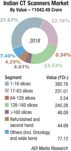
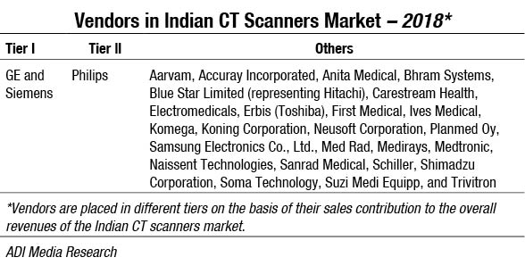
The Indian CT scanners market in 2018 is estimated at Rs 1042.49 crore, translating to 755 units in quantity terms. The 1–16 slices category dominated with a 38 percent market share. The other popular categories were 17–63 slices and 128–160 slices at a 23 percent share each. Refurbished machines continued to take a hit and were a mere 4-percent share in 2018. A large vendor as Cura is no longer aggressive. Sanrad too faced a major decline in demand. With the government directive that machines older than ten years will have to be retired has impacted sales. The market is dominated by GE and Siemens, with Philips a close second.
2019 seems to be set for a degrowth. 1H2019 had sales of 295 units, valued at Rs 369.34 crore as against 755 units, valued at Rs 524.29 crore in 1H2018, a 37 percent decline in value terms. One of the major contributors in 2018 has been a 22 units order received by GE from HLL. The government procurement was low as it was an election year, and buying decisions were in limbo at some states. The creeping in of recession, and MRI gradually being the preferred modality, may have contributed to the downward trend too.
 Global market
The global CT scanners market is estimated to rise from over USD 5.2 billion in 2018 to USD 7 billion by 2025, according to Global Market Insights. Rising prevalence of cardiovascular diseases globally will drive the market over the next 6 years. CT scanners offer multiple benefits in diagnosis and treatment of CVD that will stimulate their demand. Also, scans generated through cone beam allow healthcare providers to obtain images of heart valves and arteries that accurately predict blockage, increasing success rates of surgeries. Moreover, increasing awareness about early disease diagnosis in emerging economies will further accelerate demand for CT scanners.
Technological advancements in portable CT scanners and addition of slices will fuel industry growth. The equipment delivers superior image quality as compared to other imaging modalities. Furthermore, advancements in technology have reduced the risk associated with radiation exposure without hampering the quality of results that accelerate the demand for CT scanners. However, availability of refurbished CT scanners and high cost associated with CT scanners may impede their demand and hamper the CT scanners market growth.
O-arm segment held 60.4 percent revenue share in 2018 and is estimated to dominate the CT scanners market owing to extensive usage of O-arm in diagnosis of several chronic conditions. It is widely preferred for assessing pedicle screw position in spine surgeries and kyphoplasty procedures. Moreover, O-arm is a cone-beam imaging system that is highly preferred in minimally invasive vascular surgeries.
Stationary CT scanners market is expected to have 5.4 percent growth during 2019 to 2025 with the adoption of stationary CT scanners in hospitals that carry out large numbers of scans every year. Well-established hospitals find it convenient to assemble stationary CT scanners that have exceptional capacity and efficiency though they occupy larger space. Also, in developed countries, public hospitals are forced by the regional government authorities to adopt stationary CT scanners as results generated by these scanners are more reliable as compared to portable CT scanners that stimulate the segmental growth.
 Mid-slice segment was valued at USD 1.4 billion in 2018, and it will grow considerably with the availability of CT scanners that deliver mid-slice assortment of images, enabling efficient surgical treatment. Mid-slice technology includes scanners with 16–40 slices that possess considerable diagnostic accuracy with minimum exposure to radiations that substantially boost its adoption. Moreover, frequent launch and approval of advanced CT scanners that deliver mid-slice range will fuel segment growth.
Hospital segment is expected to show 5.3 percent growth during 2019 to 2025. CT scanners are used in disease diagnosis, treatment, and also for studying disease progression. They also provide guidance during surgery and help locating appropriate parts for operation, thereby preserving delicate structures. Therefore, hospitals have high adoption of technologically advanced CT scanners.
The CT scanners market in Germany held 23 percent of the European industry revenue share in 2018, and country growth is driven by technological advancements in portable CT scanners and high adoption of CT scanners in mobile stroke units in the country. Furthermore, significant R&D investment to develop superior CT scanning equipment with enhanced applications will augment industry size in the future. Moreover, German scientists have developed an innovative new scanner to revolutionize cancer diagnosis. The newly developed technology can give resolution of 100 nanometers, making the examination quicker and exposing patients to less radiation. Therefore, efforts in R&D will favor the German market growth.
Mexican market was valued at USD 34.8 million in 2018. Adoption of cone-beam technology for disease diagnosis is increasing steadily that should augment the country growth. CT scanners market is still in nascent stage in Mexico. Hence, new entrants have ample opportunities to grab the market share by penetrating with low-cost CT scanners.
Some of the notable global industry players are Canon Medical Systems Corporation (Toshiba), CurveBeam, Hitachi Medical Corporation, Koninklijke Philips, Neusoft, Samsung, Shenzhen Anke High-Tech, Xoran Technologies, United Imaging, Siemens Healthineers, Planmed Oy, Medtronic, Koning Corporation, GE Healthcare, Carestream, and Accuray. Industry players are focusing on R&D activities to develop innovative product delivering superior image quality in less cost.
Major players – An update
In February 2019, at the European Congress of Radiology (ECR), Philips introduced its Incisive CT scanner that will help imaging departments and healthcare organizations to meet their clinical, operational, and financial goals. The company claims that the Incisive CT scanner is integrated with several innovations in imaging, workflow, and life-cycle management.
In September 2018, GE Healthcare introduced its Cardiographe CT scanner, particularly for the coronary imaging market, by adding several new technologies that are specifically aimed for improving coronary CT angiography and radiology imaging. The scanner was developed by Israeli start-up company Arineta who took the development clearance from the US Food and Drug Administration (FDA) in 2017.
In September 2018, Siemens Healthineers shared that Ohio State University Wexner Medical Center in Columbus had installed its Somatom go.Top CT system. The Somatom go.Top CT works on the patient-centric mobile workflow concept, which is controlled through tablet and remote. This innovation addresses new researches in clinical fields and applications, especially in the field of cardiology.
Technology trends
Artificial Intelligence. A team of bioengineers from Rensselaer Polytechnic Institute (RPI) has developed an artificial intelligence (AI) technique, which converts the low-dose CT images into superior quality. In this technique bioengineers use image post-processing to convert the low-dose CT scans into superior-quality images, which are helpful in clinical research and patient care.
There are also the development algorithms that might extract the information from CT and MRI scans, which help in patient management.
Fractional-flow reserve CT. One of the new add-on advanced visualization technologies for CCTA that vendors are now offering is fractional-flow reserve computed tomography (FFR-CT). It offers a completely noninvasive FFR assessment of not just a coronary segment, but the entire coronary tree. Invasive catheter-based FFR uses blood flow pressure readings before and after a coronary lesion to determine if a stenosis is hemodynamically significant and requires stenting. Traditionally, CTA has been great at showing anatomical detail, including the percentage obstruction a lesion presents. But in the cath lab, it is often the case where a significant blockage seen on CT does not greatly impact blood flow. To avoid the need for a diagnostic catheterization for confirmation, FFR-CT uses super-computing computational fluid dynamics to assess a virtual FFR, which studies have shown has an 80-percent correlation with invasive FFR. This may be the key in making CT a true gatekeeper to the cath lab and for EDs to more quickly discharge chest pain patients where coronary blockages can be ruled out. Several vendors have recently created partnerships with HeartFlow, which offers FFR-CT technology that is cleared by the FDA.
Photon-counting CT. Photon-counting CT is an upcoming technology in the field of CT scanners with the ability to change and bring something new in the clinical CT. The photon-counting CT uses a new type of energy-resolving X-ray detectors, which is different from other conventional energy-integrating detectors.
hMachine learning. Machine learning has the potential to vastly advance medical imaging, particularly CT scanning, by reducing radiation exposure and improving image quality. Those new research findings were just published in Nature Machine Intelligence by engineers at Rensselaer Polytechnic Institute and radiologists at Massachusetts General Hospital and Harvard Medical School. According to the research team, the results published in this high-impact journal make a strong case for harnessing the power of artificial intelligence to improve low-dose CT scans.
“Radiation dose has been a significant issue for patients undergoing CT scans. Our machine learning technique is superior, or, at the very least, comparable to the iterative techniques used in this study for enabling low-radiation dose CT,” said Ge Wang, an endowed chair professor of biomedical engineering at Rensselaer, and a corresponding author on this paper, “It is a high-level conclusion that carries a powerful message. It’s time for machine learning to rapidly take off and, hopefully, take over.”
Low-dose CT imaging techniques have been a significant focus over the past several years in an effort to alleviate concerns about patient exposure to X-ray radiation associated with widely used CT scans. However, decreasing radiation can decrease image quality. To solve that, engineers worldwide have designed iterative reconstruction techniques to help sift through and remove interferences from CT images. The problem, Wang said, is that those algorithms sometimes remove useful information or falsely alter the image. The team set out to address this persistent challenge using a machine learning framework. Specifically, they developed a dedicated deep-neural network and compared their best results to the best of what three major commercial CT scanners could produce with iterative reconstruction techniques. The researchers were looking to determine how the performance of their deep-learning approach compared to the selected representative iterative algorithms currently being used clinically. This research by Wang’s team is among the significant advancements consistently being made by faculty in the Biomedical Imaging Center within the Center for Biotechnology and Interdisciplinary Studies (CBIS) at Rensselaer.
Outlook
Over the last few years, CT imaging has demonstrated continuous progress in hardware, software, and machine-learning applications. For CT hardware, significant advances have been made in scanner gantry rotation speeds improving temporal resolution, spatial resolution, detector coverage, and, therefore, overall scan times and needed contrast. In the next decades, one can anticipate continued progress in all these areas, and particularly in machine-learning applications in cardiac CT, as they are incorporated into clinical routine for image acquisition, image analysis, and pre-diction of patient outcomes.
Second Opinion
AI – The second set of eyes!
 Dr Anurag Yadav
Sr Consultant Radiologist, Department of CT & MRI,
Sir Ganga Ram HospitalAdvances in diagnostic radiology in the last two decades were primarily about improvement of hardware with more powerful and refined magnets and faster CT scanners. Software upgrades although significant, have become more important and relevant in the last few years. Imaging has developed over time from the realm of structural depiction of patient pathology to encompass illustration of tissue microstructure, physiology, and metabolism. Advances in vendor software have increased the use of advanced tools, such as MR perfusion and MR spectroscopy, diffusion tensor imaging in day-to-day practice.
Radiologists now face the dilemma of having a great wealth of diagnostic information at their fingertips, some of which is too vast to sift through, and some which they are unable to appreciate the significance of. Artificial intelligence and machine learning have stepped in to help them deal with the load of clinical work and also to try to find patterns in disease they may have been oblivious to, or detect voxel-based data alterations of disease before they are visible to the naked eye. Assigning mundane or tedious time-consuming tasks, such as cardiac segmentation and volumetry in cardiac imaging to an algorithm, will allow radiologists to spend more time on more important things that make a difference to clinical outcomes.
AI is evolving to be a second set of eyes, which learns on the job. Imagine AI and machine learning algorithms have been developed that can segregate the large numbers of radiographs at teaching institutions around the world into critical, urgent, and normal groups, and flagging emergent conditions so that they can be reviewed and informed immediately to the treating clinician.
In the world of cancer detection and treatment, AI stands to enhance screening programs and ensure treatment protocols as per the most current guidelines, the goal being uniform world-class standard of care irrespective of which hospital.
Although none of the above algorithms is perfect or currently in routine use, except for a few institutions, this is the direction we are headed in. Eventually, we expect a bundled AI and machine learning algorithm on each of our reporting systems. It will enhance and simplify work while learning on the job from each radiologist so that we may each have a virtual radiology assistant.
Technololgy trends
 Dr Mona Bhatia
Director and Head Department of Radiodiagnosis & Imaging,
Fortis Escorts Heart InstituteTechnological advancements in imaging have always made a huge difference to patient management through early diagnosis and treatment planning with accurate prognosis. While the belief was that CT advancements with increased slices and speed of acquisition had reached their peak, the end of the road with nothing more to offer, the rise in dual energy and spectral CT are demonstrating new vistas, in previously unchartered territories with revelations which will translate spectral CT to play the role of the game changer over the next decade.
Dual energy and spectral CT can differentiate tissues and pathologies based on their energy-related attenuation characteristics or material density. Three different techniques are used by different vendors for spectral CT acquisition and reconstruction including dual-source dual-energy CT, fast KV switching spectral CT, and dual-layer spectral detector CT. CT images can subsequently be used to create any desired spectral reconstruction known as spectral derived images. Virtual mono-energetic, virtual non-contrast and different chemical composition like calcium-only, iodine-only, and water-only images are easily obtained on single clicks. Hence even a single acquisition, suffices to achieve non-contrast, contrast images with tissue differentiation at low radiation dose with higher soft-tissue contrast.
Specifically, spectral CT is being increasingly used in vascular applications, where it serves as a problem-solving tool. Images can be acquired at much lower contrast dose, particularly in patients with renal insufficiency, creating virtual arterial phase images. Spectral CT in oncology is growing rapidly and is gaining ground in lesion detection, lesion differentiation between hyper and hypo vascular lesions, for follow up after therapy with the potential to eliminate need for a separate unenhanced phase acquisition of imaging, thereby decreasing radiation dose.Dual-energy CT with spectral imaging is also useful in genitourinary applications for stone characterization, renal mass characterization, tumor perfusion, and response to therapy, urothelial tumour detection, adrenal mass characterization, and artefact reduction. Neuroimaging applications of spectral CT include improved grey white matter differentiation, vascular visualization, plaque characterization, assessing hemorrhage versus calcium in non-contrast CT and iodine versus hemorrhage in post-contrast CT with analysis of underlying pathology of hematoma. In addition, spectral CT is extremely helpful in artefact reduction and bone removal in CT angiography.
Musculoskeletal applications of spectral CT include metal artefacts subtraction besides evaluation of tendons and ligaments integrity. Another critical role of spectral CT is in cardiac imaging where CT enables not only exceptional coronary assessments but superior plaque characterization and myocardial perfusion imaging with reduced artefacts making CT the only one stop shop solution for cardiac imaging. Thus, the disruptive technology of spectral CT is in fact a boon in disguise, as it improves tissue characterization with reduction in artefacts and radiation dose levels. Hence it would not be incorrect to call spectral CT the game changer of the future.


