


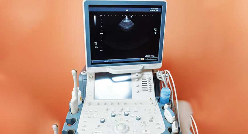
Ultrasound Equipment
Ultrasound | The evolving ultrasound market is poised to push growth boundaries
Ultrasound continues to be a game-changer, pioneering innovation and transforming medical imaging landscapes.

Continuous innovation in ultrasound equipment has propelled ultrasound technology into new frontiers, enabling healthcare professionals to explore unprecedented depths in imaging and diagnostics.
There is unprecedented clarity, more portability and versatility. The new ultrasound technology allows for greater flexibility and convenience. Advancements in signal processing algorithms are revolutionizing the way ultrasound scans are conducted.
There are now the exciting possibilities of 3D and 4D ultrasound imaging, offering a new dimension to the field. And the integration of AI algorithms identify subtle abnormalities that may be challenging to detect manually.
Pioneering on an unstoppable path of innovation, medical imaging technology has reached new heights, with ultrasound equipment leading the charge.
Significant advancements
Advancements in technology have revolutionized healthcare by allowing doctors to accurately diagnose conditions and plan appropriate treatment options.
Improved image quality and resolution. Visualization of intricate structures with exceptional clarity is now possible, facilitating the detection of even the smallest abnormalities in organs and developing fetuses. Radiologists and sonographers can confidently identify subtle changes in tissue texture.
Enhanced portability and versatility. More compact and lightweight devices facilitate easy transportation and use across various healthcare settings. It enables medical professionals to swiftly assess patients at their bedside, in remote locations, or during emergencies, leading to expedited diagnoses and improved treatment outcomes.
Enhanced versatility, allowing for a broader range of diagnostic procedures within a single device is offered. From obstetrics and gynecology to cardiology and musculoskeletal imaging, healthcare professionals can seamlessly transition between different imaging modes, adjust settings, and even perform real-time 3D imaging for comprehensive evaluations.
Signal processing algorithms have revolutionized ultrasound technology by significantly enhancing the quality and clarity of images, leading to more precise diagnoses and treatment plans. They enable ultrasound machines to detect and enhance subtle details, reduce noise and artifacts, and improve visualization of specific structures or organs.
3D and 4D ultrasound imaging gives an extraordinary level of detail and depth, allowing real-time visualization of unborn babies’ movements and features. It provides both a captivating visual experience for expectant parents and valuable medical information for healthcare professionals. And enables accurate measurement, monitoring of growth and development, and early detection of potential abnormalities or complications, significantly enhancing prenatal care and facilitating early interventions if needed. Such ultrasound imaging revolutionize obstetric medicine, providing unprecedented insight into prenatal healthcare.
Improved techniques
The recent strides in ultrasound have had transformative effects on bedside patient care. Some techniques that have paved the way for enhanced patient care include:
Elastography measures tissue stiffness, very valuable in differentiating between benign and malignant masses, as tumors.
Elastography and shear wave imaging are useful for assessing liver stiffness, which correlates with the grade of fibrosis, the degree of portal hypertension, and the risk of developing hepatocellular carcinoma. These imaging modalities provide non-invasive methods to evaluate liver health and are particularly useful for diagnosing liver fibrosis and cirrhosis, reducing the need for invasive procedures like liver biopsy.
Shear wave elastography, a specific form of elastography including both 2-D shear wave elastography and point-shear wave elastography, has high sensitivity and specificity in detecting liver fibrosis with advantages over transient elastography in terms of accuracy and the ability to assess liver stiffness with high precision.
Additionally, they can be used as predictors for the severity of fibrosis, potentially reducing the necessity for liver biopsy in patients with chronic liver disease.
Fusion imaging combines ultrasound with other imaging modalities as CT or MRI. This integration allows for the overlay of ultrasound images onto the anatomical context provided by other imaging techniques. Fusion imaging improves accuracy during image-guided procedures and facilitates better localization of lesions such as hepatocellular carcinoma and prostate cancer. By combining contrast-enhanced ultrasound with subsequent tomography, image fusion facilitates improved detection and characterization of liver lesions, leading to better treatment outcomes.
Additionally, image fusion enhances biopsy techniques, such as in the case of prostate cancer detection, by allowing for targeted biopsies based on fused MRI and ultrasound datasets. This approach not only increases the detection rate of significant cancers but also reduces the risk of overtreatment by minimizing the detection of clinically non-significant cancers.
What lies ahead
Nomogram combines ultrasound features to assess thyroid nodules. Researchers led by Lina Pang from Fourth Military Medical University in Xi’an, China, have developed a nomogram that combines ultrasound features with thyroid imaging reporting and data system (TI-RADS) parameters to differentiate between malignant and benign thyroid nodules. The study demonstrates the high predictive performance of the nomogram, particularly for TI-RADS categories 3, 4, and 5.
Ultrafast technology. A research team led by Professor Chulhong Kim and colleagues from various departments and institutions in South Korea has developed a technique for imaging the three-dimensional microvasculature of the kidneys using ultrafast ultrasound. Their method, which does not require contrast agents, has been recognized for its ability to visualize the entire kidney microvasculature.
This achievement is significant for understanding kidney function and diagnosing conditions like kidney failure, especially in patients with hypertension and diabetes. By capturing 1,000 frames per second, the researchers were able to image the renal vascular network, including arteries and veins, as well as interlobular vessels, with unprecedented detail. Their findings, published in Advanced Science, demonstrate the potential of this technique for studying vascular changes in kidney diseases and monitoring outcomes of kidney transplantation.
Wearable ultrasound scanner. Researchers at MIT, led by associate professor Canan Dagdeviren, have developed a wearable ultrasound device designed to detect breast tumors in their early stages.
The team, including lead authors Wenya Du, Lin Zhang, Emma Suh, and collaborators from Xi’an Technological University, designed a miniaturized ultrasound scanner based on piezoelectric materials, enabling portability and ease of use. Testing the device on a human subject, the researchers successfully detected breast cysts as small as 0.3 centimeters in diameter, comparable to early-stage tumors.
AI in ultrasound. AI and machine learning algorithms are being incorporated into ultrasound systems to automate image analysis, optimize image quality, and assist in diagnosis. AI algorithms can aid in the detection of abnormalities, reduce operator dependence, and improve overall workflow efficiency.
AI in obstetrics and gynecology ultrasound imaging has the potential to reduce inter- and intra-observer variability by automating image acquisition and interpretation processes, thus minimizing clinician workload, and improving diagnostic accuracy.
It also benefits patients by reducing unnecessary hospital admissions and interventions. It supports clinical education by providing skill training for novices and enabling non-experts to acquire ultrasound images, during times of increased workload or shortages of experienced sonographers.
However, limitations exist, such as the need for human expertise in image acquisition and interpretation, the dependence on specific gestational age ranges for biometric measurements, and challenges related to image artifacts and noise. Ethical considerations, including concerns about algorithm safety, trustworthiness, and privacy, also warrant attention.
The integration of AI algorithms into fetal ultrasound systems diminish the reliance on operator expertise. It can streamline workflow processes by automating routine tasks and providing real-time assistance and such AI-enabled software can assist in image optimization, fetal biometry measurements, and anomaly detection, thereby enhancing efficiency and productivity during prenatal examinations.
Outlook
The year 2023 marked a significant leap forward in ultrasound technology, characterized by exciting advancements across various fronts. With unprecedented clarity and portability, modern ultrasound systems transcend boundaries, enabling healthcare professionals to delve deeper into imaging and diagnostics. From 3D and 4D imaging to AI integration, each stride forward brings new vistas of possibility, enhancing our understanding and treatment of various medical conditions.
In the pursuit of early detection and personalized medicine, ultrasound’s role is paramount. From detecting breast tumors with wearable devices to targeting Parkinson’s disease with focused ultrasound, each breakthrough paves the way for improved patient outcomes and a brighter future in healthcare.


