
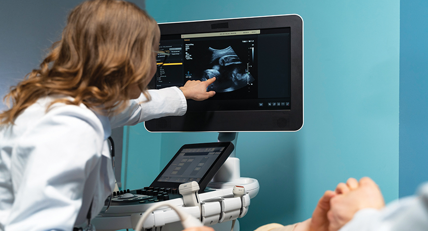
Ultrasound Equipment
Revolutionizing accessibility and convenience with ultrasound
The transformative metamorphosis is ushering in a new era, where ultrasound is not merely a diagnostic tool but a therapeutic force to be reckoned with.

In the ever-evolving landscape of medical technology, the resounding pulse of progress beats through the corridors of ultrasound innovation, creating not a mere ripple but a seismic shift. Recent times have witnessed a surge in the prominence of ultrasound equipment, propelled by remarkable technological advancements and ground-breaking developments. As the industry invests fervently in the orchestration of research and development (R&D), the trajectory of ultrasound technology promises a meteoric rise, leaving an indelible mark on the future of healthcare.
This transformative metamorphosis is propelled by an unyielding commitment to pushing the boundaries of possibility, ushering in a new era, where ultrasound is not merely a diagnostic tool but a therapeutic force to be reckoned with.
As the resonance of ultrasound technology reverberates through the medical landscape, its impact extends beyond clinics and hospitals, woven into the very fabric of healthcare delivery. The future, pulsating with anticipation, holds the promise of remarkable strides, poised to transcend the current impact and shape the healthcare landscape in unprecedented ways. Applications of ultrasound, from guiding minimally invasive procedures to revolutionizing therapeutic interventions, are expanding at a revolutionary pace.
The future heralds not only improved image quality and resolution but also a significant leap in portability and versatility for ultrasound machines. No longer confined to specific settings, these portable machines provide healthcare professionals with valuable diagnostic tools, wherever they are needed, revolutionizing accessibility and convenience.
Yet, the symphony of progress extends further. Advancements in signal-processing algorithms revolutionize the conduct of ultrasound scans, enhancing accuracy and efficiency for quicker and more reliable results, ultimately elevating patient care standards. Exciting possibilities in 3D and 4D ultrasound imaging open new dimensions, allowing healthcare professionals to visualize and analyze the human body in unprecedented ways.
Indian market dynamics
The Indian ultrasound market in 2023 is estimated at ₹1845 crore, by value, and 10,600 units. Despite a distinct preference for high-end models, as no new models were introduced nor any distinct features added, it is the mid-end models that saw an appreciable increase. The portable segment continues to cater to niche customers. The refurbished segment contributed 7 percent to the market and is gradually losing share.
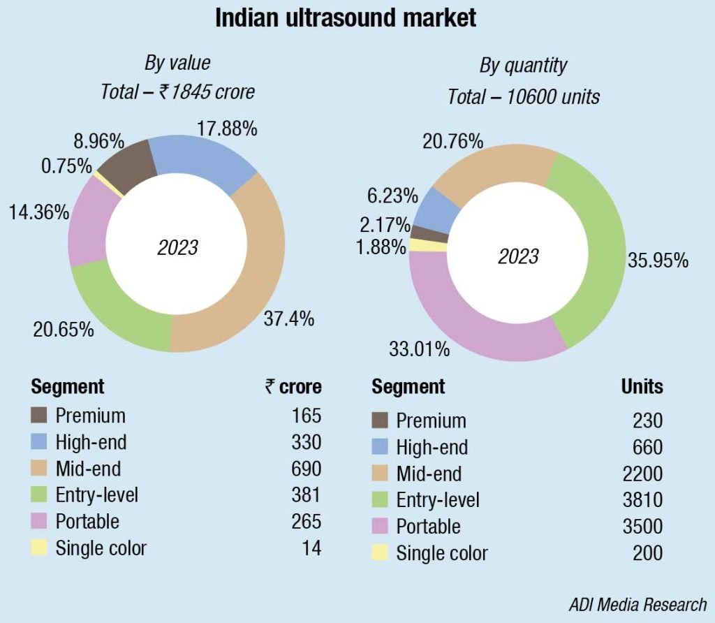
By type, the cart/trolley-based machines continued to dominate with 81.4-percent share, and the balance 18.6 percent was contributed by the compact machines.
GE continues to dominate this market with a huge lead. Samsung and Mindray are competing for the second slot, though the Korean brand remains ahead of its Chinese counterpart. Philips occupies the third position.
The larger hospitals are replacing their machines within 2–3 years as against 5 years earlier, and in the smaller cities within 5 years. This has helped expand the size of the market.
The vendors in turn are offering discounts and rewarding loyalty. A couple of vendors are not offering exchange schemes, letting the machines remain with the hospital, in case of repeat buying.
|
Leading players – Indian ultrasound market-2023 |
|
| Tier 1 | GE |
| Tier 2 | Mindray, Samsung & Philips |
| Tier 3 | Sonosite & BPL |
| Tier 4 | SonoScape & Esaote |
| Others | Trivitron, Konica, Toshiba, Aloka, Toshiba, and Cura |
| *Vendors are placed in different tiers on the basis of their sales contribution to the overall revenues of the Indian ultrasound market.
ADI Media Research |
|
North India dominated the market in 2023, particularly cities like Delhi, NCR, and Chandigarh, which have a high concentration of well-established hospitals, healthcare facilities, and medical institutions, making them primary consumers of ultrasound systems, according to a Tech Sci Research study. Due to higher population densities, greater healthcare spending capacity and a rise in medical colleges, research institutions are more likely to adopt the latest medical technologies, which include modern ultrasound systems.
Affordability is a concern. With out-of-pocket health expenditure expenses accounting for about 63 percent of total health expenditure, one of the highest in the world, high costs deter individuals from seeking necessary diagnostic services. In rural and remote areas of India, healthcare facilities may lack the necessary infrastructure and resources to acquire and maintain advanced ultrasound equipment. This leads to disparity in healthcare access. Operating ultrasound equipment requires trained and skilled personnel, including radiologists and sonographers. The shortage of such professionals in some areas can limit the accessibility of ultrasound services.
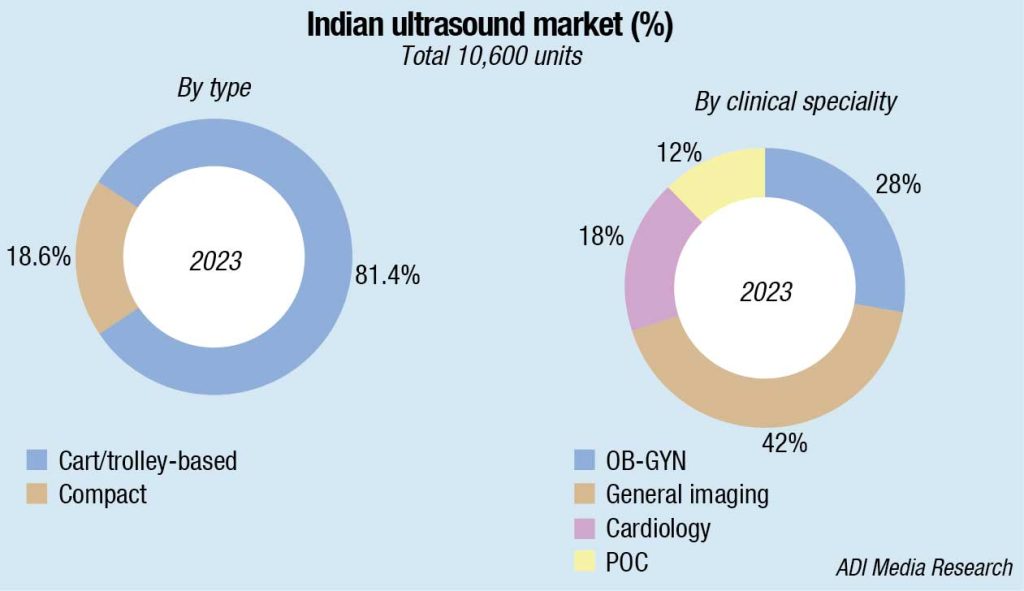
The diagnostic segment accounts for the largest share. This is owing to the wide range of applications in radiology, cardiology, obstetrics, gynecology, internal medicine, and more. Point of care in hospitals and clinics allows immediate and on-site assessment of various medical conditions demanding diagnostic ultrasound. These systems are often more cost-effective than highly specialized ultrasound equipment and are, therefore, a more attractive option for healthcare institutions.
Technological advancements and emerging trends on ultrasound technology
 Preeti Vimal
Product & Application Manager-Premium Ultrasound,
BPL Medical Technologies
In the ever-evolving field of medical diagnostics, imaging technologies, particularly ultrasound, have undergone remarkable transformations. The continuous innovation has propelled ultrasound technology into new frontiers, enabling healthcare professionals to explore unprecedented depths in imaging and diagnostics. As we enter a new era of healthcare, the continuous advancements in ultrasound equipment are shaping the future of non-invasive medical examinations, presenting more opportunities for an improved patient care.
An exciting advancement in ultrasound technology is the integration of artificial intelligence (AI). By incorporating AI algorithms into ultrasound equipment, healthcare professionals are now equipped with a powerful tool for analyzing the images. This not only aids in faster and more accurate diagnoses but also assists in identifying subtle abnormalities that may be challenging to detect manually. The amalgamation of human expertise and AI capabilities holds immense opportunity in medical diagnostics.
The rise of three-dimensional (3D) and four-dimensional (4D) imaging technologies are also gaining prominence. These innovations allow for a more comprehensive visualization of the patient’s internal structures, offering a more detailed and realistic representation of anatomical features. This has proven particularly valuable in areas, such as obstetrics, cardiology, and musculoskeletal imaging applications, where precise visualization is vital for diagnoses and further treatment planning.
Advancements in ultrasound probes and signal-processing techniques enable improved image resolution, image contrast, and clear details, which in-turn increases the operator’s ability to detect subtle abnormalities. Keeping the unique challenges of the Indian healthcare landscape into consideration, portable and handheld ultrasound devices emerge as invaluable upgradation. Their compact nature and ease of portability facilitate increased accessibility and flexibility, allowing healthcare practitioners to conduct ultrasound examinations at the point of care. This is particularly beneficial in remote or resource-limited settings, where traditional machines may be impractical to install.
Ultimately, ultrasound technology will reach its full potential through a collaborative ecosystem comprising of healthcare providers, researchers, experts, and partners ready to create equipment with the latest features. The continuous evolution of ultrasound technology paints a promising picture for the future of even more advanced and personalized healthcare solutions that ultimately benefit patients as well as healthcare providers.
The general imaging segment has the largest share and will continue to expand in the coming years. Such ultrasound is often used for routine health checkups and screening, often at the point of care in both hospitals and clinics, for quick and early assessment of various medical conditions and due to being cost-effective, increasing its market share.
Global market dynamics
The global medical ultrasound market in 2023 is estimated at USD 8.5 billion, expected to grow to USD 12–13 billion by 2030, a CAGR of 5 percent. Six players dominate the segment. GE dominates with a 29 percent share, Philips contributes 19 percent, Toshiba- Canon 11 percent, Mindray, Fujifilm-Hitachi, Siemens, Fujifilm-Sonosite and Samsung Medison each are in the 5-8 percent range, Esaote, BK Medical, SonoScape, and many other brands together constitute the balance 9 percent.
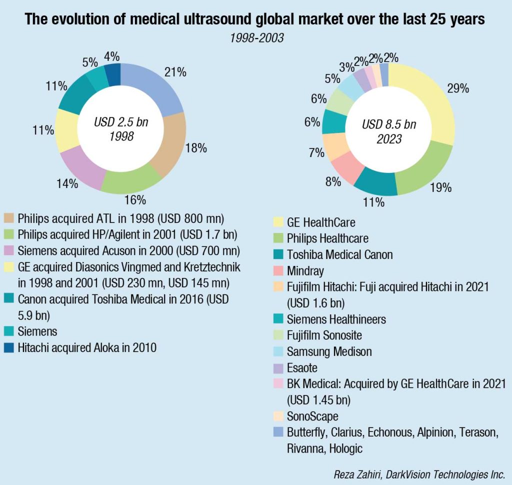
Ultrasound imaging has been one of the fastest growing sectors compared to other imaging modalities. Over the last 25 years, medical ultrasound imaging market has grown from USD 2.5 billion in 1998, to USD 8.5 billion in 2023 worldwide, a CAGR of over 5 percent. Reza Zahiri, PhD MBA, VP Research & Innovation, DarkVision Technolgies Inc., has very accurately tracked the development.
Writes Zahiri,“The high growth rate made the ultrasound market very attractive. This is evident by looking at the history of the acquisitions especially in late 1990s and early 2000s.
1998 – Philips acquired ATL Ultrasound (market leader, 18 percent market share).
1998 – GE acquired Diasonics Vingmed.
2000 – Siemens acquired Acuson (14 percent market share).
2001 – Philips acquired Agilent Healthcare/HP medical (16 percent market share).
2001 – GE acquired Kretztechnik from Medison.
The trend continued:
2010 – Samsung acquired Medison.
2010 – Hitachi acquired Aloka.
2012 – Fuji acquired Sonosite.
2016 – Canon acquired Toshiba Medical.
2018 – A Chinese consortium acquired Esaote.
2021 – GE acquired BK Medical.
2021 – Fuji acquired Hitachi Diagnostics Imaging.
As the ultrasound market grew, the premium that big companies are willing to pay for gaining more market share has also increased:
1998 – Philips paid USD 0.8 billion for 18 percent market share (ATL acquisition).
2012 – Fuji paid USD 1 billion for 6 percent market share (Sonosite acquisition).
2021 – GE paid USD 1.45 billion for 2 percent market share (BK Medical acquisition).”
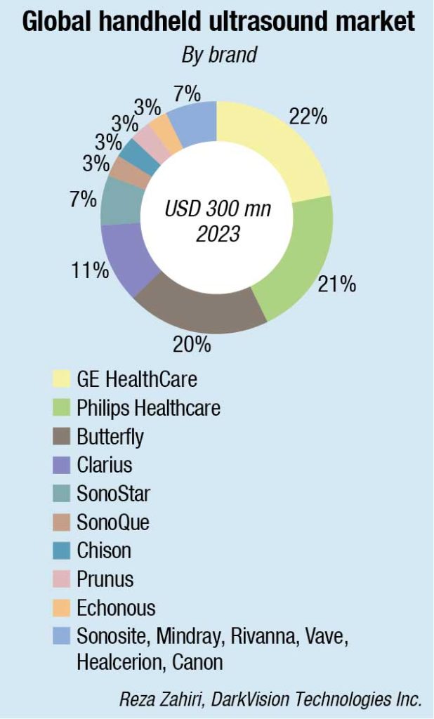
Over the past eight years, handheld ultrasound systems have become the fastest-growing segment in the ultrasound market, estimated to reach USD 2.5 billion by 2026. Handheld devices also remove several barriers to entry. Many handheld systems cost a small fraction of the cost of the laptop and cart-based systems, representing 60–90 percent savings. They are easier to use, take up very little space, and can be carried in a pocket. Many handheld ultrasound devices also provide comparable image quality to most mid-range traditional ultrasound systems. As older systems need to be replaced, many specialists are switching to high-definition handheld options for their practice, including plastic surgeons, orthopedic surgeons, and physiatrists who prefer a more portable wireless system.
Point-of-care ultrasound (PoCUS) includes the use of smaller, less expensive ultrasound systems to assess, triage patients in an office, or emergency department setting. The imaging modality offers immediate images with no waiting to assess, if a patient requires immediate change in care or more advanced imaging assessment. PoCUS is also used in catheterization labs to guide vascular access and to guide anesthesia needle placement. PoCUS systems also are being used in developing countries as a way to access immediate, inexpensive medical imaging.
Technological advancements
Ongoing advancements in ultrasound technology have driven the adoption of newer ultrasound systems.
Ultrasound transducers have advanced to include higher-frequency probes, which results in improved image resolution. This allows for more detailed and clearer images, especially in superficial structures.
Capacitive micromachined ultrasonic transducer (cMUT) technology. The progress in cMUT design and operation has led to improved performance, and the technology holds promise for developing lower-cost, 3D imaging devices with integrated on-chip electronics. Recent releases of commercial probes, utilizing cMUT technology, indicate the beginning of realization of this promise. These probes are being increasingly used in various clinical ultrasound applications. As cMUT technology continues to establish itself in the landscape of ultrasound transducers, it is expected to have a growing impact, offering cost-effectiveness and enhanced imaging capabilities for years to come.
Ultrasound localization microscopy. ULM is an emerging technique showing promise in providing super-resolved images of microvasculature for both preclinical and clinical applications. It offers improved resolution compared to traditional ultrasound techniques, such as contrast-enhanced ultrasound (CEUS) and Doppler methods, allowing imaging and flow measurements down to the capillary level. ULM relies on localizing single microbubbles (MB) of contrast agents, enabling sub-pixel precision imaging and the tracking of MBs for both morphological and functional information, including flow velocities and directions. The method has demonstrated potential in various applications, including assessing pathological and physiological changes in the microvasculature.
The transition to clinical applications is an ongoing area of research, with considerations for imaging in 3D to compensate for tissue motion. Current clinical applications work with available acquisition modes and DICOM data, limiting the method’s accuracy. The future of ULM in clinical settings depends on manufacturers implementing ULM on their systems and developing dedicated imaging modes.
Noncontact laser ultrasound (NCLUS). Researchers from MIT Lincoln Laboratory and Massachusetts General Hospital have developed noncontact laser ultrasound (NCLUS), a laser-based ultrasound system for medical imaging. This laser-based ultrasound system provides images of interior body features, such as organs, fat, muscle, tendons, and blood vessels. The system also measures bone strength and may have the potential to track disease stages over time.
Addressing limitations of traditional contact probes in ultrasound, NCLUS aims to automate the imaging process, reduce operator variability, and provide a fixed-reference frame. The system uses a pulsed laser to generate ultrasound waves through a thermoelastic process, offering noncontact measurement without tissue distortion. The clinical NCLUS system features miniaturized lasers, a portable armature, and faster imaging capabilities, making it suitable for various applications, including military and civilian healthcare settings. The technology’s potential for automation and portability positions it as a transformative tool, comparable to MRI and CT.
The team is now developing NCLUS to support field-forward military applications. These applications include detecting and characterizing life-threatening injuries from internal bleeding in organs; monitoring debilitating musculoskeletal injuries, and their healing over time; and providing elastographic imagery of soft tissue and bone of amputee limb regions to accelerate the design and fitting of prosthetic sockets. Civilian applications include imaging in the intensive care unit. With NCLUS, emergency medical technicians, paramedics, and medical staff without specialized sonography training might be able to perform ultrasound imaging outside of a hospital – in a doctor’s office, at home, or in a remote battlefield setting.
While it may not yet be categorized as a widespread technology trend, NCLUS demonstrates characteristics of an emerging technology with transformative potential. Its ability to provide automated, portable ultrasound imaging with a fixed-reference frame aligns with the ongoing trend of advancing medical technologies for improved accuracy, accessibility, and efficiency. Once clinical studies are successful, the team will seek commercial funding for clinical medical devices development, followed by US FDA approval.
3D ultrasound tomography. A novel 3D ultrasound tomographic (3D UT) method (called volography) that creates a speed of sound (SOS) map and a reflection modality that is co-registered are reviewed and shown to be artifact-free even in the presence of high contrast and thus shown to be applicable for breast, orthopedic, and pediatric clinical use cases. The 3D UT images are almost isotropic with millimeter resolution and the reflection image is compounded over 360 degrees to create sub-millimeter resolution in plane.
Ultrasonic system-on-patch (USoP). Researchers at the University of California, San Diego (UCSD), have developed the ultrasonic system-on-patch (USoP), a stick-on ultrasound patch, representing the first fully integrated wearable system for evaluating cardiovascular function during activities like walking, running, or cycling. The USoP combines an ultrasonic sensor and a miniaturized flexible control circuit in a single wireless wearable device, eliminating the need for cables for both power and data transmission, and enhancing mobility. The device incorporates a machine-learning algorithm that automatically analyzes signals and instructs the ultrasonic sensor to track targeted tissues as they move, and then interprets incoming data by using advanced machine learning.
By offering real-time data for enhanced patient outcomes, the USoP exemplifies how the Internet of Medical Things (IoMT) transforms patient monitoring. This medical device can be used in various medical settings, including emergency medicine, primary care, and remote areas, potentially improving patient care by providing continuous monitoring of vital signs, which can help detect health issues early and improve patient outcomes.
New ultrasound method could lead to easier disease diagnosis. Researcher Dr Artur Gower from the University of Sheffield, in collaboration with Harvard, Tsinghua University, and the University of Galway, has developed a breakthrough ultrasound technique to measure tension in soft tissue. Unlike traditional ultrasounds, this method utilizes sound waves to measure tension, allowing for better diagnosis of abnormal tissue, scarring, and diseases, such as cancer. The technique, based on the principle that greater tension results in faster sound wave propagation, is the first capable of measuring tension in any type of soft tissue without prior knowledge. The breakthrough could be used to build new ultrasound machines that are able to better diagnose abnormal tissue, scarring, and cancer.
AI integration
Even with handheld systems, more work is required to lower the learning curve for novice users to use ultrasound imaging. Even advanced users require time and attention to optimize the image, which takes their eyeballs off the patient. That is why artificial imaging (AI) has become the key to unlocking the tremendous potential of handheld ultrasound by automatically optimizing image quality and guiding the users on probe positioning. For example, a handheld ultrasound device can use AI to automatically capture high-quality images of every body part. A clinician simply places the scanner on part of the abdomen, and the app automatically detects the organ and optimizes the image without intervention. Research finds that using ultrasound with AI capabilities can increase practitioner accuracy and reduce false positives by 37.3 percent.
Way forward
Ultrasound technology will reach its full potential through an ecosystem of partners, ready to create solutions and applications for their specialties. Ultrasound will also reach its full potential faster as vendors use open platforms that allow for the integration of third-party innovations. Focusing more on complete solutions and less on standalone hardware will allow ultrasound innovators to lower the barrier to entry around education and image acquisition, interpretation, and procedural guidance.
Future innovations in ultrasound plus technology hold promising possibilities. From artificial intelligence integration to the development of miniaturized devices, the field of ultrasound is poised to continue evolving and revolutionizing medical imaging. The future of ultrasound technology is bright, and its potential to improve healthcare outcomes is truly remarkable.
Second Opinion:-


