


MB Stories
Expanding the possibilities of an established technology
Despite its age, x-ray imaging remains a versatile technology that continues to find innovative applications in diverse fields. The integration of AI algorithms has paved the way for automated image analysis, enabling faster and more accurate diagnosis while minimizing human error.

The advent of diagnostic medical imaging can be traced back to the introduction of the x-ray unit by Roentgen in 1895. x-ray technology has revolutionized the field of medical diagnostics since its discovery over a century ago.
Since its inception, radiographic imaging has undergone significant advancements, revolutionizing the field of diagnostics and treatment over the last century. Traditional x-rays, the earliest form of diagnostic imaging, have paved the way for the development of more sophisticated technologies, such as computed tomography. Initially employed for imaging skeletal structures, x-ray equipment has continually evolved, expanding its applications to encompass a broad range of medical and non-medical domains.
The development of x-ray equipment paved the way for unprecedented insights into the human body, enabling healthcare professionals to detect and diagnose various ailments with greater precision and efficiency. From identifying fractures and tumors to visualizing internal organs and monitoring treatment effectiveness, x-ray imaging has become an indispensable tool in modern medicine. Its ability to provide real-time, high-resolution images, coupled with its non-invasive nature, has significantly contributed to enhancing patient care and reducing the need for more invasive procedures.
The remarkable versatility and non-invasive nature of x-rays have propelled their integration into numerous fields, revolutionizing patient care, scientific research, and industrial processes.
Indian market dynamics
The Indian x-ray market in 2022 is estimated at ₹1000 crore by value and 16850 units by numbers. There is a distinct shift from analog systems to DR models.

Within the DR segment, the imported systems dominate with 85-percent share by quantity. And the DR retrofit detectors are the popular machines. The full room DR x-rays are finding value from the discerning customers, although they are priced above ₹7,500,000.
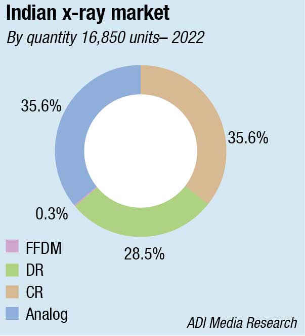
The CR systems have a 36-percent share by quantity, albeit only an 18-percent share by value.
In April 2023, the government had hiked customs duty on import of x-ray machines and non-portable x-ray generators from 10 percent to 15 percent. The changes in the customs duty rate were brought in as part of the amendments to the Finance Bill, 2023, which was passed by the Lok Sabha. And it brought all the x-ray components under the same harmonization code. This, accompanied by a further weakening of the rupee, resulted in an increase in prices of the machines.
|
Leading players* Indian analog market |
|
| Tier I | Allengers |
| Tier II | Vision, Kiran, Medion & Epsilon |
| Others | Skanray, BPL and regional fragmented brands |
|
Indian CR market |
|
| Tier I | Fujifilm |
| Tier II | Agfa, Konica and Carestream |
|
Imported DR fixed – Full room |
|
| Imported | Agfa, Siemens, Philips, Fujifilm, Carestream, & Konica, Shimadzu, and Samsung |
| Indian | Allengers, Skanray & United Imaging |
|
Indian DR mobile market |
|
|
Imported brands |
|
| Tier I | Samsung |
| Tier II | Carestream |
| Others | Agfa |
| Indigenous brands | |
| Tier I | Allengers |
| Tier II | BPL |
| Others | Skanray & Kiran |
| Indian retrofit detectors market | |
| Tier I | Konica, Fujifilm, CareRay and Carestream |
| Tier II | Agfa, and PZ Medical |
| Indian FFDM market | |
| Imported brands | |
| Tier I | Fujifilm and Siemens |
| Tier II | Hologic, GE, and United Imaging |
| Indigenous | Allengers |
| Indian DR OEM panels market | |
| Tier I | iRay |
| Tier II | Varian & DR Tech |
| Others | PerkinElmer, Varex, Toshiba, & Thales |
| *Vendors are placed in different tiers on the basis of their sales contribution to the overall revenues of the Indian x-ray market.
ADI Media Research |
|
The industry is finding regulatory compliance a huge challenge. Presence of multiple regulators makes simple tasks, such as rectification of an erroneous declaration on the label, quite a drawn-out process. While CDSCO regulates medical devices, quality management system requirements and Essential Principles of Safety & Performance for Medical Devices are defined by BIS, the diagnostic facility is licenced by AERB, the National Pharmaceutical Pricing Authority fixes prices, legal metrology rules, Department of Pharmaceuticals, and state FDAs also regulate the industry. Despite all this, spurious films and other components are being sold at lower prices with barely any penalties, leaving the safety of the patients compromised. The presence of laws that restrict manufacturers and importers of medical devices from promoting their products directly to the customers in certain circumstances also poses a challenge.
The introduction of PACS and digital patient records has not eliminated the demand for diagnostic hardcopy. One particular aspect that deserves mention is the use of non-tested, non-approved paper interfaced with standard printers, resulting in inaccurate diagnosis of x-rays, CT, or MRI.
Bridging the gap with affordable digital radiography solutions in India
 Roy Sebastian
NPI Leader – Imaging,
Skanray Technologies Limited
Digital radiography has revolutionized the healthcare domain, offering numerous advantages over film-based and computerized radiography systems. India recognizes the need for cost-effective digital radiography solutions.
Cost efficiency
Replacing film-based and computerized radiography systems with digital radiography significantly reduces costs. Conventional methods involve expenses for film, chemicals, and maintenance. Digital radiography eliminates these costs, leading to substantial long-term savings. Affordable digital radiography solutions enable Indian healthcare institutions to allocate resources effectively, improving patient care and outcomes.
Enhanced workflow and efficiency
Digital radiography accelerates the entire imaging process, enabling immediate image acquisition, review, and transfer, and allows healthcare professionals to capture images instantly. Moreover, digital images can be easily shared and accessed by multiple specialists, promoting collaboration and enabling timely diagnoses. These advancements enhance workflow efficiency, reduce waiting times, and ultimately contribute to better patient management.
Improved diagnostic accuracy
The ability to enhance and manipulate digital images enables radiologists to detect subtle abnormalities more accurately. Digital images can be magnified and adjusted for optimal visualization, aiding in the identification of intricate details. Embracing affordable digital radiography solutions bridges the gap between urban and rural healthcare facilities in India.
Reduced environmental impact
Digital radiography supports sustainable healthcare practices. Unlike film-based systems, digital radiography reduces the environmental impact of Indian medical institutions. Electronic storage of digital images eliminates physical storage and reduces paper consumption. This shift showcases the healthcare sector’s commitment to sustainable development.
Outlook
Affordable digital radiography can transform Indian healthcare by streamlining imaging, enhancing accuracy, and reducing costs. Digital radiography also supports global sustainability efforts. Collaboration among policymakers, healthcare institutions, and technology providers is vital to ensure affordable digital radiography throughout India, improving patient care and efficiency.
While many printing technologies are available today, not all comply with the quality standards required to produce a diagnostic-quality image on a transparent diagnostic film. Direct thermal printers and printer systems can deliver the image quality required for diagnosis.
Contrast resolution, spatial resolution, and optimal viewing conditions are key parameters that define diagnostic quality, but the physical properties of the material are also important. Paper/opaque printing systems offer a lesser degree of value for diagnosis due to limited contrast resolution and use of half-tone printing.
The manufacturers have brought these issues to the attention of the powers-that-be under the umbrella of associations as FICCI, and are awaiting action.
Global market scenario
The global medical x-ray market is estimated at USD 13 billion in 2022 and is poised to reach USD 23 billion, at a record 5.9-percent CAGR from 2023 to 2032. North America is the largest market, expected to be USD 7.3 billion by 2032.
The increasing prominence of diagnostic imaging procedures, such as x-ray examination procedures mainly amongst the geriatric population, are expected to counter the rising risk of chronic diseases and complement the medical x-ray industry progress. The surging awareness of early disease diagnosis has also proved favorable for the rising number of diagnostic procedures. The continuous advancements in medical imaging techniques, including computed tomography (CT) have further resulted in the rapid demand for x-ray procedures, further influencing the market revenue.
The declining patient preference for x-rays, considering their risks of harming the body cells, may pose as a constraint for the industry progress. x-rays have high energy as compared to light waves, and thus offer high-energy radiation, subsequently increasing the burden of cancer. Hence the rising number of performed x-ray tests may hinder the market share. x-rays also lead to ionizing radiations, resulting in pre-and postnatal irradiation, bringing adverse health impacts for children.
Within the segment, medical x-ray market from the fixed portability segment is slated to record USD 12.5 billion by 2032. The rising number of government investments for installing modern diagnostic equipment have spurred the adoption of fixed x-ray systems as they can be accessed in a wide variety of sizes and types in several high-end scientific applications. These systems also address the imaging needs across all clinical settings. The rising popularity of mammograms and dental x-rays for facilitating the imaging of skull, chest, extremities, spine, and abdomen will also add to the market outlook.
Medical x-ray market share from chest applications is poised to record over USD 6 billion by 2032 due to the rising need to produce images of bones of chest, airways, spine, blood vessels, and lungs. Chest x-rays are used for diagnosing as well as monitoring the treatment of lung cancer, pneumonia, emphysema, heart problems, fractures, heart failure, etc. It also looks at the placement of medical devices, such as catheters, pacemakers and defibrillators.
The thriving requirement for detecting symptoms, including fever, breathing difficulties, persistent cough, and chest injuries will also accelerate the demand for chest x-rays.The rising popularity of detector-based digital radiography x-ray is accelerating industry growth. x-ray detectors are easing the healthcare professionals’ work in the diagnosis and further treatment of patients. The effective use of advanced equipment, such as x-ray detectors, helps to screen a larger number of patients at lower costs and is time-saving. Growing support, funds, and investments will greatly drive access to the adoption of these technologies, which can help the market to grow further.
Estimated to be worth USD 3.1 billion in 2022, it is poised to reach USD 4.0 billion by 2027, growing at a CAGR of 5.2 percent from 2022 to 2027. The x-ray detector market is constrained by the high cost of these devices. This is not only the constraining element, it also makes it challenging for poor and growing nations to adopt new technologies. Many hospitals also have insufficient funding, thus they are unable to even consider purchasing these modern technologies. Security checks are no longer done manually as every nation strives to develop and adopt cutting-edge technologies. Then there is the idea of a health hazard; in security applications, these detectors have been shown to induce cancer, which can be a real limitation. Another issue that needs to be addressed is the scarcity of qualified radiologists.
In the x-ray detectors market, emerging nations like APAC and India have many prospects. These countries are seeing a large growth in medical tourism, thanks to factors, such as the expanding population, rising sedentary lifestyles, and the quick growth of medical tourism. These factors are contributing to the rise in health conditions, such as cardiovascular and neurovascular diseases. Additionally, these nations have returns on investments that are better than average, and are thought to have a skilled labor force. These countries’ medical tourism industries are being nurtured and grown by government programs and policies.
Major players in the x-ray devices and equipment market are Siemens Healthcare, Carestream Health, Philips Healthcare, Hitachi Medical, GE Healthcare, Toshiba Medical Systems Corporation, Canon, Siemens Healthcare, Philips Healthcare, GE Healthcare, Fujifilm Holdings Corporation, and Shimadzu Corporation.
The development of digital x-ray devices has opened up new possibilities for utilizing this technology in various physical settings and disciplines. The lightweight, wireless, and robust cassette-sized detector, used in DR technology, which can transform analog x-rays into complete digital radiography solutions, makes it an effective technology. With the use of this technology, digital x-ray images are produced, which eliminates the need for film or computed radiography (CR) plates.
In addition, compared to its digital counterpart, film-based traditional radiography requires more manpower, and even the consumables utilized are more expensive. Chronic diseases, such as cancer, cardiovascular, COPD, CKD, and diabetes, are the major causes of death among adults in developing as well as developed countries. According to the WHO, chronic disorders account for about 41 million deaths globally, which is equivalent to 74 percent of all fatalities worldwide. Digital x-ray technology helps in the early intervention of such disorders, thus boosting its demand. Moreover, compared to traditional x-rays, this technology provides a number of benefits including 70 percent less radiation, better image quality, and less acquisition time.
The global digital x-ray devices market size is anticipated to reach USD 4.5 billion by 2030, according to a recent report by Grand View Research, Inc., anticipated to expand at a CAGR of 3 percent. Factors, such as the increasing prevalence of chronic disorders, rapid technological advancements, and increasing patient understanding of early diagnostic techniques, are all augmenting the industry’s growth. The fixed digital x-ray segment accounted for the largest revenue share in 2022 owing to the introduction of technologically advanced devices, while the mobile digital x-ray segment is expected to register the fastest CAGR.
New innovations drive the market
CXLS. Arizona State University, USA, has commissioned a first-of-its-kind instrument that will help scientists see deeper into matter and living things. The device, called the compact x-ray light source (CXLS), generated its first x-rays on the night of February 2, 2023, by ASU scientists. The CXLS provides hard x-ray pulses with high flux, stability and ultrashort durations, in a very compact footprint. This way, matter can be resolved at its fundamental scales in space and time, enabling new discoveries across many fields – from next-generation materials for computing and information science, to renewable energy, biomolecular dynamics, drug discovery and human health.
Scintillators, which have been in use for around 70 years, have undergone improvements to enhance their emission brightness and speed. A new design, representing a new approach to fabricating high-performance x-ray imaging scintillators based on organic metal halides for applications in medical radiography and security screening, has been developed by a team of scientists in Saudi Arabia. Herein, the zero-dimensional organic copper halide (18-crown-6)2Na2(H2O)3Cu4I6 (CNCI), which exhibits negligible self-absorption and near-unity green-light emission was successfully deployed into x-ray imaging scintillators with outstanding x-ray sensitivity and imaging resolution. In particular, they fabricated a CNCI/polymer composite scintillator with an ultrahigh light yield of ~109,000 photons/MeV, representing one of the highest values reported so far for scintillation materials. In addition, an ultralow detection limit of 59.4 nGy/s was achieved, which is approximately 92 times lower than the dosage for a standard medical examination. Moreover, the spatial imaging resolution of the CNCI scintillator was further improved by using a silicon template due to the wave-guiding of light through CNCI-filled pores.
A team of four from the Israel Institute of Technology has authored a paper, published in Advanced Optical Materials, using the concept of inverse-designed nanophotonic scintillators, which can overcome the trade-off between resolution and efficiency by reshaping the intrinsic spontaneous emission. To exemplify the concept, multilayer phosphor nanostructures are designed, and these nanostructures are compared to state-of-the-art phosphor screens in image intensifiers, showing a threefold resolution enhancement, simultaneous with a threefold efficiency enhancement. The enabling concept is applying the ubiquitous Purcell effect for the first time in a new context – for improving image resolution. This approach directly applies to a wide range of technologies, including x-ray imaging applications.
Furthermore, the manipulation of length scale and optical properties of scintillators has led to the creation of nanophotonic scintillators. Researchers have successfully calculated the scintillation levels produced by various nanophotonic structures. The European Synchrotron Radiation Facility’s Extremely Brilliant Source (EBS) project has utilized these advancements to achieve non-destructive, three-dimensional (3D) scans of intact human organs at the cellular level. This breakthrough enables high-resolution imaging without the need for stains or contrast materials, reducing dose exposure and improving image quality.
Maximum radiation protection & minimum patient dose
 Ramesh Chandra Satyawali
National Sales Head, DR and X-ray Business, Healthcare Division,
FUJIFILM India Private Limited
In the era of a constantly evolving digital epoch, radiation technology has progressed tremendously. Dose reduction and radiation protection areas have a huge scope for improvement and integration of new-age technologies in mobile x-ray systems. This is foremost important for enhanced diagnosis, minimum side effects and safety of patients and healthcare professionals.
How mobile X-rays can limit the radiation dose
The significance of state-of-the-art mobile x-rays can be derived from the fact that they ensure the lowest possible radiation dose to the patient. A multitude of technological inclusions in this type of x-ray allows the portable x-ray technology to emit the lowest amount of radiation, thereby, ensuring protection from radiation.
High-frequency (HF) x-ray generator technology. HF x-ray generators are designed to efficiently generate, and control high-voltage power required to drive the x-ray tube, while also being user-friendly. They are efficient and capable of managing 40 percent less radiation dose, and have superior image quality. Therefore, these generators not only provide top-quality imaging but also prioritize patient safety.
Collimators. Collimators are used to confine the x-ray beams to the body part of the patient required to scan, preventing unnecessary radiation exposure to other parts of the body. A good collimator system in mobile x-ray machines offers a precise field view along with increased radiation safety.
Highly sensitive flat-panel detector. Effective mobile x-ray machines comprise of highly sensitive flat-panel detectors (FPD), which produce sharp and clear images at low radiation, compared to traditional flat-panel detectors. In addition, some highly-sensitive FDPs consist of unique noise-reduction circuit. This unique technology serves best when integrated with advanced light signal-capturing techniques within the FPD. ISS is one such technology.
Grid-less Image Acquisition Software (Virtual Grid). It is the avant-garde of image-processing software technology that rectifies the effects of scattered radiation, which otherwise reduces image contrast and clarity. Furthermore, it eliminates the requirement for an anti-scatter grid and quickly produces high-quality images, thereby resulting in reduced radiation dose to the patient, yet superior image quality. It is a relatively novel innovation in image-processing software technology that uses an algorithm instead of a physical grid.
The combination of nanophotonic and scintillation technologies has the potential to further enhance resolution, reduce x-ray dose, and facilitate energy-resolved x-ray imaging. Moreover, the integration of HiP-CT with clinical images, using AI techniques, can validate ambiguous findings with high accuracy and improve information deduced from other imaging modalities.
HiP-CT technique. The HiP-CT technique has shown promising results during the global pandemic, offering insights into the impact of Covid-19 on blood oxygenation. By bridging the gap between CT and MRI scans and histology, HiP-CT allows researchers to visualize small vessels, specific cells, and microscopic structures within organs. This has provided direct evidence of the lung abnormalities associated with Covid-19, such as shunting.
The data obtained from HiP-CT, along with molecular methods, has aided in understanding the pathophysiology of Covid-19 and its effects on oxygen levels and the circulatory system. These advancements have paved the way for hierarchical 3D imaging of multiple intact human organs, contributing to the development of The Human Atlas and Human Biomolecular Atlas, which aim to map the hierarchical structure of human organs.
Overall, these advancements in x-ray imaging technologies hold great potential for delivering better patient care by enabling more accurate diagnosis, faster imaging, reduced radiation exposure, and improved understanding of health and disease.
Spectroscopic x-ray imaging. Eleven years ago, there was a lot of skepticism about the technical feasibility and the clinical benefits of spectroscopic x-ray imaging. The sixth Workshop on Medical Applications of Spectroscopic x-ray Detectors at Cern in August 2022, celebrated a major milestone for the technology, the US Food and Drug Administration (FDA) approval of the first new major technological improvement for computed tomography imaging in nearly a decade in the form of a photon counting scanner. This is the first scanner using spectroscopic x-ray imaging officially approved for regular medical use in the world. Spectroscopic x-ray imaging is now set to revolutionize diagnostic medical imaging by providing better images with a lower dose to the patient, allowing new workflows that optimize precious hospital resources. When spectroscopic detectors are used, the images also contain the colors of the incoming x-rays, providing better, clearer images at optimized doses with significant benefits, when diagnosing disease. In some circumstances, MRI may even become superfluous. In other cases, where metal contrast agents attached to bio-markers are injected into the body, expensive PET-CT scans, may be avoided. Spectroscopic detectors yield much more information than DECT systems. A single image is taken. No upfront decision is needed and no extra dose is used – the energy information is always available.
Some medical and industrial x-ray imaging applications need to reconstruct an image on a flexible surface, so they use photographic film rather than electronic detectors. Current flat-panel x-ray imaging detectors are difficult to adapt to these applications. In the FleX-RAY project, flexible x-ray imaging detectors using scintillating fibers have been used. The FleX-RAY research project demonstrated it to the scientific community at the 24th International Workshop on Radiation Imaging Detectors, held in Oslo. FleX-RAY uses a sheet of flexible scintillating fibers to detect x-rays and guide the scintillation light to arrays of silicon photomultipliers. The detector also self-reports its curved shape, using optical waveguides with Bragg gratings in a flexible glass substrate, which act as curvature sensors. Multiple reconstruction algorithms have been developed, suitable for different x-ray energies.
Recent imaging breakthroughs and future directions
Some recent x-ray imaging developments are set to change the face of radiography in the next few years. These include:
Flexible x-ray imaging. Up until recently, most x-ray detector panels were flat and only capable of scanning x-rays in a 2D format. A large rotating gantry has allowed for 3D imaging to take place. However, due to the expense and equipment size, inventors have been looking for 3D imaging alternatives. One such alternative would be flexible imaging panels that curve around surfaces. In the last few years, several flexible panels have been invented that improve upon all currently available flat panels (direct and indirect types) used in medical imaging devices. Some of these new inventions can curve around surfaces and may even be worn by the patient for comfortable scanning of selective body regions. Unfortunately, these prototypes contain heavy metals, such as lead, which is likely to increase any potential radio-toxic side effects.
Liquid nanocrystal imaging. In 2021,liquid nanocrystals were discovered that were capable of capturing x-rays better than currently available flat panel technology, allowing for optimal flexible x-ray imaging without heavy metal involvement. This technology also solves problems pertaining to irregular shapes, which often show up blurred in conventional radiographs. Yet, these liquid crystal imaging devices are not able to convert high levels of radiation into images, suggesting the course of future innovations.
Portable x-ray imaging devices. With the invention of flexible panel detectors of all shapes and sizes, the possibility of portable x-ray devices is moving closer to becoming a reality. In tandem with flexibility, inventors have been working on making portable x-ray imaging devices and incorporating them into everyday gear, such as cellphones, cars, and glasses. As of 2022, an imaging plate, the size of a square millimeter, has been developed that can detect large quantities of photons on its minuscule surface area with a relatively high degree of accuracy. With more advancements such as these, x-ray imaging devices may become as small as thermometers and integrated into the diagnostic toolkit of any general practitioner. With more time and refinement, early detection of cancer and other tissue abnormalities could be part of an everyday standard check-up.
Improved computation. Advances in computer software have led to better imaging as well. Algorithms have been developed that improve the accuracy of x-ray-generated images, particularly through better recovery of blurred areas and other irregularities that tend to show up on the images. These improvements are more relevant to CT scanning. One recent development allows for flawless x-ray imaging of the heart by using an algorithm that incorporates the patient’s ECG results with the sensor. The scanner is able to capture more accurate x-ray images of the heart from multiple angles that are matched in time to the patient’s heartbeat, thereby enhancing the resolution of the final image.
Artificial Intelligence has shown great promise. There is an exponentially growing demand for medical imaging. Meanwhile, there is a global shortage of trained radiologists. AI methods benefit radiology practice, which ease their work like more accurate classifications, enhanced analysis, generating 3D models and quicker results. Certain challenges including lack of standardization, lack of explainability, lack of validation datasets, and breach of privacy exist. Despite all these challenges, AI helps radiologists to solve problems and have some practical applications.
One of the greater misconceptions about radiologists is that they essentially just analyze images. Such simplified assessments, dramatically oversimplify what radiologists do. The tasks a radiologist performs on a regular basis far outweigh the capacity of current technological functions. Such work includes patient-facing work to consulting with other physicians, multi-disciplinary work, training, and audits.
Current radiology AI systems perform single tasks, undertaking specific image recognition, such as nodule detection on a chest CT scan. These narrow, numerous, and necessary detection tasks are required to fully diagnose the image findings. AI can play a substantial part in improving the diagnostic workflow, even replacing a human in some of the more mundane tasks, such as scheduling. But unless we miraculously invent a complete end-to-end system that includes qualified oversight over the entire diagnostic pathway, AI will not replace radiologists entirely anytime soon.
Efficiency is of particular importance in the field of radiology, given the shortage of devoted professionals globally. AI can alert radiologists to acute conditions in patients in an efficient and timely manner, accelerating the time it takes to address cases
A transformation for the field
One of the most exciting developments we can expect is the shift from active to proactive detection of medical conditions. Rather than just looking into the specific medical condition for which the patient arrived and requested medical care, AI will empower radiologists to discover additional conditions that were previously undiagnosed or even unknown to the patient. This could lead to the discovery of vertebral compression fractures or cardiovascular events. Through the early detection of such conditions, enabled by AI, radiologists will be able to employ the right treatment for patients at risk for osteoporosis, for example, a game changer for patients and radiologists alike.
We can also look forward to the utilization of AI to extract discrete data from medical images that’s reproducible, quantifiable and extensible. Achieving exact and accurate measurements means that specific pixel data can be leveraged to unlock innovative application tools and big data analytics. Combining this with other pertinent data sources, such as genetic sequencing, will give birth to a diagnostic power play: leveraging insights to go beyond just prevention to drive customized therapeutics, and personalized care.
As AI becomes more immersed in the more laborious, time-consuming functions of radiology tasks, it will begin generating new, unanticipated results that can then be used as sources of medical discovery. Not only will this evolution move forward radiology, but a host of other medical fields and research domains will flourish.
Radiologists won’t be replaced. However, by embracing AI and adapting to these changing times, they will see their jobs transformed and their patients’ quality of care improve. Aided by AI, the field will continue to thrive.
The global AI in medical imaging market size is expected to reach USD 8.18 billion by 2030, based on a recent report by Grand View Research, Inc. The market is expected to expand at a CAGR of 34.7 percent from 2022 to 2030 owing to the growing aging population and increased investment in the healthcare ecosystem by the government and private players.
The increased adoption of interventional x-ray equipment, such as C-arms, for image-guided surgeries is the key factor driving the segment.
High demand for minimally invasive procedures, a well-established health sector, broadening clinical uses for x-ray detectors, and rising demand for early diagnosis will keep this segment going for years to come.
Current scenario and trends in x-ray market
 Dr Madhur Saxena
Senior Consultant Radiologist,
Bhagwan Mahaveer Cancer Hospital, Jaipur
While talking about current scenario and trends in x-ray market, first thing I would like to comment upon is that though it is most basic and ancient radiological investigation, yet wide-spectrum of its utility, instant availability, and cost effectiveness will never let it go outdated. Either preventive or therapeutic diagnostics, role of x-rays is stupendous in both the applications.
As we peep into gradual technical improvement from conventional radiography to CR and DR systems, we have not only enhanced image quality and post-exposure modification options but have also significantly improved timings of entire procedure and radiation doses. Besides improving quality to detect smallest possible abnormalities, we are focusing on reducing doses also to attenuate radiation exposure. Low-dose, high-quality radiography systems are the demand of the market.
Further, new-generation advanced technologies have opened an entire new chapter in radiography, converting grey scale images to revolutionary colored radiographs (transmutation from boring black and white images to interesting colored images). Inverted images are also available to make any particular pathology more obvious and prominent.
Role of radiographs has also been extended beyond imagination as per the demands of the clinical domain. A few latest uses of radiographs to be noted are:
- Carotid artery atherosclerotic plaque calcification detection serves for prevention of ischemic cerebral stroke, which in most of the cases is result of extra-cranial atherosclerosis. Dedicated software and hardware can detect and measure calcific plaques in carotid vessels in the neck.
- Automated cardio-thoracic ratio calculation. cardiomegaly is detected automatically through analysis of digital fluorograms, thus allowing identification of patients at risk of cardiac diseases.
- Osteoporosis screening. Identification of patients at risk for osteoporosis and bone fractures is of utmost importance. The latest available x-ray technologies can identify subset of patients who should be referred for further clinical assessment with DEXA.
- Image optimization. Optimum visualization of both high-density and low-density pathologies in single exposure.
Another interesting area, I would like to discuss is portable use of radiography machine. Though portable x-rays are being used in clinical set-ups since long but these are the x-rays in which many challenges are being faced by the medical staff. Be it improper positioning due to lack of patient compliance in ICUs, motion artifacts, breathing artifacts, countless lines and tubes, or unnecessary radiation exposure to all people present in ICU, the list of challenges is inexhaustible.
Better technical expertise of radiation staff, advanced machines (low dose – high SNR), and patience and clinical correlation while reporting are some of the solutions I can think of.
To reduce unwanted radiation exposure to rest all people in wards or ICUs, now digital chest screening units are also available. These units provide closed chambers to expose patients while saving all others from radiation.
CT x-ray systems are also available as mobile devices for instant bedside imaging of patients. These are compact systems with open bore and low radiation doses to patients. These machines can be operated at standardized voltage, so can be used anywhere.
In conclusion, I would like to again emphasize that though the latest USG, CT scan, and MRI systems play tangible role in diagnostic field, yet the role of x-ray system should not be undervalued, and we all should hopefully look forward for consistent improvements and technical advancements to further enhance its utility.


