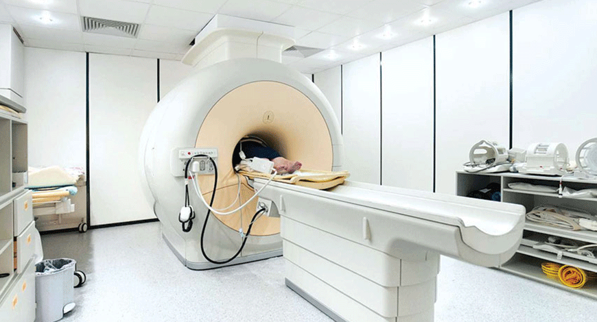
MRI Equipment
Game changing innovations on the horizon

MRI technology has attained a push toward greater evolution rates because of a deep interest shown by manufacturers and technology developers. Improvements in the technology are working in favor of the global MRI market.
Advancements in technology pertaining to magnetic resonance imaging (MRI) have enabled capturing of high-quality images of ligaments, soft tissues, and other body organs. Moreover, technological advancements have widened the scope of applications. Companies are focusing toward research and development of advanced open systems, which have minimum chances of suffocation and higher acceptance among claustrophobic people.
Hence, such improvements in the technology are working in favor of the global MRI market. The rising geriatric population is also stimulating the adoption of MRI.
The demand for MRI is strong for scanning brain and neurological systems due to the lack of effective and accurate alternation. However, in several other applications such as cardiac and breast imaging, alternatives such as ultrasound techniques and X-ray are considered to be first-line imaging tools. In terms of field strength, mid field strength devices are likely to have high demand over the next 5 years, owing to affordable price and acceptable speed and resolution.
The global MRI market is expected to be dominated by international players with their strong foothold and brand name across all major geographies. A large number of players will be focusing toward product innovations to stay relevant in the market.
By 2024, the total MRI global market is estimated to garner revenue of USD 4.52 billion from USD 3.99 billion in 2019, up at a compound annual growth rate (CAGR) of 2.5 percent, estimates Frost & Sullivan. However, with the COVID-19 outbreak, the market experienced a slowdown and is likely to reach the pre-COVID levels in 2023, registering impressive growth.
Growing public-private partnerships and an increase in near-replacement time in public domains are expected to attract more MRI system procurements. Technologies such as point-of-care, pediatric, dry-magnet, and compact MRI and fusion imaging are driving the global MRI market.
The open MRI segment will continue to diminish with wider adoption of 1.5T and 3T MRI systems that incorporate increased field of view and radiofrequency (RF) channels, and enhanced signal-to-noise ratio, optimizing patient throughput with improved static and dynamic imaging capabilities and artificial intelligence (AI)-augmented technical features. The commercialization of portable MRI is expected to increase dramatically in the mid to long term. The inclusion of technologies that improve workflow, portability, and the ability to cater to various applications are competitive factors driving the market.
From a market segment perspective, high-field and extremity MRI are vital contributors to the total MRI market, garnering revenue at a CAGR of 8.5 percent. In developing economies with a large population base, improved healthcare facilities and physicians’ preference for 1.5 T MRI is preferred over other conventional medical imaging methods.
The key driver for the global advances in MRI market currently is the increase in healthcare expenditure that is being funneled into research and development activities. Additionally, physicians are shown to hold a greater favoring toward modern MRI systems over traditional medical imaging modalities, further improving the chances of research and development activities in the global MRI market. Private hospitals are expected to lead the demand for MRI devices, and the adoption rate of MRIs is likely to grow because of the growing occurrences of cardiac diseases, neurological, and oncological cases.
The challenge faced by the global advances in MRI market is the probable rise in the prices of hospital-grade helium. Liquid helium is used for lowering the temperature of superconducting magnets in MR imaging systems. Severe shortage of liquid helium is expected to limit the market growth as it will affect the rate of production for OEMs in it.
MRI technology has attained a push toward greater evolution rates because of a deep interest shown by manufacturers and technology developers. These developers and manufacturers wish to make it more patient-friendly in light of the improving standards of patient satisfaction and positive recordkeeping.
The last few years have seen significant advances in the field of MRI imaging. Some of the most important upgrades include:
MRI-conditional implant scans. Medical implant technology has grown increasingly sophisticated, but implants traditionally complicated the MRI scanning procedure. Nowadays, however, spinal implants, pacemakers, and other devices are becoming MRI-conditional, which means they do not interfere with the scanning process within certain parameters. In the past, the scanner needed to be manually adjusted to budget for the presence of the implant. But Philips recently unveiled an MRI automated user interface model which patients to be scanned faster and easier.
Silent scanning. Noises during spinal imaging can be a real headache. With the new SilentScan package included in GE Healthcare’s Signa Pioneer 3T system, the noise created during spinal imaging and other types of MRI processes can be greatly reduced. This system includes an Ambient Experience technology package that offers a soothing experience for the patient complete with imagery, sound and light specially designed to help patients relax. Ingenia also offers ScanWise implant technology, which greatly simplifies the process of scanning patients with spine implants and other types of implants.
High-resolution images: Higher magnetic field strengths of the newer MRI machines ensure very high quality images compared to the lower magnetic fields of the traditional MRI machines. However, the 1.5T MRI machine is considered ideal for most MRI procedures. It can create better images than the lower strength 1.2T MRI machines, and because of the variety of coil options and established protocols can potentially produce better images than a 3T MRI machine. From a cost standpoint, a 1.5T MRI scan also costs half as much as a 3T MRI scan.
Faster procedure. The advances in technology and magnetic field strengths have shortened the amount of time the patient has to lie still in the magnet for the procedure, which could be up to an hour or longer with older MRI machines. At Houston MRI, the average time spent in the magnet is about 15-20 minutes.
Comfortable experience. When patients are comfortable during the procedure they will be able to lie still and this improves the quality of the images. The advanced short bore MRI systems are about 50 percent shorter than traditional MRI systems and also wider ensuring a more comfortable patient experience.
Abbreviated protocols. The future of breast screening may not be mammography but MRI. One of the biggest advancements in MRI breast imaging has been the creation of abbreviated protocols that can cut down breast scan time to mere minutes. Abbreviated MRI was first described in a 2014 study when researchers from Germany demonstrated how a protocol that only included the most essential precontrast images and one postcontrast image could cut breast scan time to just three minutes. Since then, numerous studies have confirmed abbreviated MRI’s diagnostic accuracy and have explored its potential as a screening method. One of the latest studies on abbreviated MRI showed that a 10-minute MRI scan found more than twice the number of invasive cancers in women with dense tissue than digital breast tomosynthesis (DBT). The same research team is now investigating whether abbreviated MRI screening could be cost-effective and still accurate if conducted every two to three years, according to Partridge.
Diffusion-weighted imaging. Aside from the time and cost, one of the biggest drawbacks of MRI is the need for gadolinium. Consequently, the panelists repeatedly lauded diffusion-weighted imaging (DWI) as one of the promising methods for contrast-free MRI. Researchers still have not eliminated the need for gadolinium contrast, which adds to both the time and potential toxicities of the exam. Diffusion DWI holds the greatest potential of a noncontrast imaging technique. On DWI, breast malignancies often exhibit more restricted diffusion and lower apparent diffusion coefficient (ADC) values than benign lesions. Early research has shown a cutoff ADC value of 1.53 x 10⁻³ mm2/sec or less could reduce biopsy rates by more than 20 percent without missing cancers.
MR spectroscopy. One of the more unconventional ideas presented at the SMRT 2020 panel was the use of MR spectroscopy to predict the development of invasive breast cancer. In her presentation, Carolyn Mountford, PhD, a professor of radiology at the University of Queensland, described how her research team has edged closer to such a technology over the past 30-plus years. “The goal of this program is an imaging test for diagnosis of early changes to breast tissue prior to life-threatening cancer,” she said.
In 1984, Mountford discovered she could use long T2 resonance to distinguish between malignancies that were metastatic and not metastatic in rats. Through a series of experiments, she realized that MR spectroscopy was picking up on specific chemical changes that predated cancerous tumors turning invasive. “We found that during the preinvasive stages of tumor development, significant changes were taking place in the plasma membrane,” she said. Thanks to data advancements, Mountford and her team utilized data mining to confirm that they could use chemistry to identify malignancy in 400 fine-needle breast biopsies. By using MR spectroscopy to just examine chemistry, the team accurately distinguished between malignant and benign tissues — as well as tissues from patients with and without lymph node involvement. “The laws of chemistry apply whether the sample is in the test tube, the membrane, the cell, the biopsy, or in vivo in humans,” she said.
Mountford is optimistic about the future of the technology, particularly for patients with very dense breast tissue. While the acquisition and processing of the data require a highly skilled radiographer or MR tech, her team is in the process of automating classifiers to evaluate the data. “It’s a new screening method for people at high risk for breast cancer potentially reducing exposure to gadolinium-based contrast agents,” she said. “It is also a personalized screening for women at average and high risk of breast cancer.”
As the demand for better diagnosis and non-invasive procedures is growing, people look forward to innovations that can enable faster contrast scans and simplify imaging workflow. An increase in investment is expected to propel the growth of the MRI market size. It is expected that the rapid development of intraoperative MRI and its use in neurosurgery and other such applications will propel the growth of the market.
Advances in diagnostic techniques, such as open MRI, visualization software, and superconducting magnets, are predicted to further fuel the MRI market in coming years. In addition, the production of cardiac pacemaker-compatible MRI systems is expected to propel the cardiology segment demand.
 Dr Harsha C Chadaga
Senior Consultant and Head,
Radiology Services, Columbia Asia Radiology Group
“With improvement in reconstruction techniques now patients with MR compatible implants can be scanned to obtain optimal images. The indications in neurological and musculoskeletal systems are ever being expanded with increasing field strength with clinical use of 7T systems. Availability of point of care and compact MRI is increasing the indications of MRI at bedside. The other areas where MRI development is making rapid strides is in dry magnet which are compact and along with fusion imaging has wider clinical applications. These are some of the incremental innovations that have improved clinical imaging. With technology developers exploring many new opportunities, especially those involving neurology, breast imaging, abdominal, and cardiac, it is inevitable that MRI would progress into a highly sophisticated medical imaging tool in the future


