
X-ray Equipment
Surgical imaging market hit by COVID-19
Demand for image-intensifier mobile C-arms was less impacted than for FPD equivalents in 2020 due to COVID-19 with reduced capital budgets, as providers focused on cost-effective equipment.

The overall economic impact of COVID-19 has been wide-ranging, affecting all industries. Medical imaging is the largest driver of the diagnostic segment of the healthcare equipment market, and has experienced a wide range of fluctuation. The pandemic has caused an unprecedented negative impact on the interventional and surgical X-ray market. During this period, most hospitals have deferred their ongoing purchase of capital equipment. A similar decrease in system installation and utilization was witnessed in 3Q and 4Q 2020, given the recurrence of COVID-19.
The interventional X-ray market has seen a higher decline in revenues than the mobile C-arm market, due to the higher associated investment required for system installations. The number of interventional procedures is expected to remain below the pre-pandemic era, thus delaying many new purchases until the pandemic subsides.
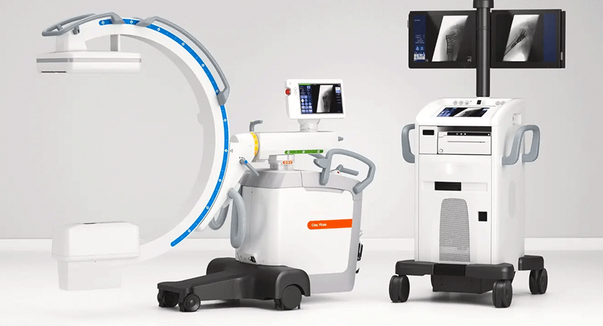
India in progressive mode with digital technology in C-arms
 Suresh Sharma
Chairman cum Managing Director,
Allengers Group of Companies
Today’s healthcare environment requires product and service solutions that can deliver and manage the ever-changing challenges of patient demographics, wherein the application of orthopedics has been fast developing with the advancement of technology.
Due to the prevalence of interventional procedures on the rise, the demand for better medical imaging is growing exponentially. The FPD C-arm segment is the major revenue contributor, and is estimated to grow significantly during the coming years because of its benefits over its counterparts. Furthermore, advantages offered by FPD C-arm over the I.I based C-arm, such as higher field of view (FOV), higher resolution, clarity, contrast, and high efficiency will further support the market demand and its subsequent growth.
FPD C-arm offers better image because of its higher dynamic range, and reduces the radiation exposure during long surgical procedures. High grey-shade levels make FPDs superior to their I.I (image intensifier)-based counterparts. More grey shades produce very-high contrast images, especially in case of uneven anatomical densities.
With the advent of newer technologies, the FPD-based C-arms are gaining momentum. From the times when hip/joint replacements, musculoskeletal repairs, ligament repairs, reconstruction to acute orthopedic surgeries were done through conventional methods of X-rays, fluorescent screens, to the image intensifiers and now to the FPD C-arms, the transition is smooth but fast. Now the surgeon, be it interventional radiologist, orthopedist, gastroenterologist, neurosurgeon, urologist, etc., can view/review his ongoing surgery through such image-guided facilities available in C-arms, empowered with dynamic flat panel detectors (FPDs ) and its advanced software. Even during the blazing pandemic in 2020, the demand for C-arms with FPDs escalated to all-time high.
The major manufacturers, who were able to visualize the possibility of such a transformational change in technology happening, were able to provide offerings that met the customer’s expectations. Now new innovations and study of C-arms for better imaging are resulting in enhanced customer experience.
As India is now going head strong with the opportunity to expedite technology adoption, as such the key take away message, is to adopt digitization, wherever possible. As the C-arm technology is re-evolving, so the companies today, more than ever, are constantly improving processes and raising standards in order to serve their customers more effectively with the finest products possible, and this is just the beginning of a new era in digital C-arms.
The challenges faced by hospitals during the COVID-19 pandemic have also severely impacted cancer care. Early in the pandemic, breast cancer patients reported significant delays in treatment, and while the number of procedures has slowly begun to return to pre-pandemic levels, the resultant backlog of diagnoses and treatments will have dangerous implications for patients for years to come. The pandemic has dramatically hindered mammography equipment sales in 2020. However, demand for interventional X-ray, mobile C-arm, and mammography X-ray systems is forecast with gradual return in 2021 and 2022, with delayed or pending orders being processed.
The global surgical-imaging market is projected to reach USD 2.4 billion by 2025 from an estimated USD 1.8 billion in 2020, at a CAGR of 6.3 percent, predicts MarketsandMarkets. In the surgical X-ray market, flat panel detectors (FPD) 2D mobile C-arms are forecast to have the fastest growth through to 2024, with a CAGR of 3.4 percent. In 2020, there was a shift toward low-end to mid-range FPD 2D systems as a result of stretched capital expenditure budgets. Demand for image-intensifier (II) systems is slowing down, with the key markets now the emerging regions. However, in developed markets, such as the United States, usage of imaging-intensifier systems is still high in pain management clinics. 3D mobile C-arms are primarily used for imaging the joints, spinal fusion, and fractures and the market is expected to start seeing signs of recovery from 2021 onwards, following the return of elective spinal procedures to pre-pandemic levels. Countries with the highest adoption of 3D surgical X-ray imaging include China, Western Europe, and the United States.
Within the interventional X-ray market, the interventional cardiology (IC) market was more negatively impacted by COVID-19 than interventional radiology (IR) due to heavier reliance on elective procedures. An increase in the number of structural heart procedures performed, in particular percutaneous coronary intervention (PCI), continues to be a factor maintaining clinical demand for the IC market. The IR market has experienced a continued expansion of clinical procedures being performed. An increased incidence of peripheral vascular disease is driving demand for general vascular angiography. Despite an estimated 26-percent drop in the hybrid operating room segment in 2020, fastest growth is predicted for this product category through to 2024.
From 2021 onwards, there is expected to be pent-up demand for both interventional and surgical X-ray systems. With the postponement of non-essential elective surgeries and medical procedures to conserve medical resources for COVID-19 patients, cardiovascular procedures were severely impacted during 2020. However, the massive financial burden of COVID-19 on healthcare providers is expected to result in reduced capital expenditure budgets for imaging equipment, including interventional X-ray and mobile C-arm systems.
While PCI procedures are assumed to be critical in treating acute myocardial infarctions (MI), only a small amount of PCI procedures performed on patients were for these life-threatening conditions. The rest of the PCI procedures are considered as elective procedures, and were deferred during the pandemic. Globally, hospitals saw a decline in cardiovascular procedures. According to a report published by researchers of CovidSurg Collaborative, around 28 million surgeries were canceled across the globe during 12 weeks of peak disruption during the COVID-19 pandemic. The American Hospital Association estimated an average loss of revenues to US hospitals of USD 50.7 billion per month from March 1 to June 30, 2020 due to loss of elective procedures.
Demand for image-intensifier mobile C-arms was less impacted than for FPD equivalents in 2020 due to COVID-19, as providers focused on cost-effective equipment due to reduced capital budgets.
The major players operating in the interventional X-ray market are GE Healthcare, Philips Healthcare, Siemens Healthcare, Technix, Hologic, Carestream Health, Canon Medical, EcoRay, Simad, Shimadzu Corporation, Toshiba Corporation, Ziehm Imaging, Hitachi Ltd., Teleflex Incorporated, and Fujifilm Corporation among other domestic and global players.
What to consider when buying C-arm?
While that might sound like a simple question on the surface, the answer is somewhat complicated. The fact is there is no one-size-fits-all solution that can be applied to every facility. Every office-based lab, clinic, and healthcare system has very specific needs that must be taken into account.
From system features to equipment size, there are strengths for each of these technologies. Some of the factors that buyers may consider are:
Dose. One of the most significant differences when it comes to FPD and image intensifiers is the radiation dose. While the technology powering both types of systems has progressed dramatically over the years, with each new iteration designed to reduce the amount of radiation, the patient and the healthcare team may be exposed to, there is a limit to what an image intensifier can accomplish. The simple fact is that flat-panel technology will almost always give you a lower dose than an image intensifier.
One can really see this come into play when working with the magnification modes available on each system. In order to get down to the Zoom 3 setting on an image intensifier, the radiologist will need to increase the dose by five times as much as they would in the normal full-field mode. The same magnification on a flat-panel detector, on the other hand, involves an increase of about half the increase. If keeping dose low is the buyer’s primary concern, then their decision is an easy one – flat panel technology. But that is only one factor of many to take into consideration.
Service and parts. Many organizations opt for the image intensifier due to an innate familiarity with the system, which manifests in easier on-site troubleshooting among the in-house team. Most C-arm operators have years of experience working with image intensifiers, so they know the quirks of the system and can pretty quickly ascertain what to do if they experience some kind of error. It is also fairly easy to find parts when a portion of the system breaks down; the widespread availability of image intensifiers means buyers are never too far from a vendor who can provide them with the part they are looking for.
Analog versus digital mammography
 Satyaki Banerjee
Satyaki Banerjee
Trivitron Healthcare
Mammography is a specialized diagnostic test carried out for the detection of early signs of breast cancer in women when there are no visible signs and symptoms of the disease. A mammogram delivers information on the morphology, normal anatomy and the gross pathology of the breast.
The radiologist conducts screening by placing the patient’s breasts on a platform. The x-ray generator modulates incoming voltage to supply the power required by the x-ray tube to generate an x-ray beam. A stream of electrons is accelerated to high velocity by a high voltage supplied by generator that collides with the tube’s target anode. As x-rays exit the tube, filters are placed through the path to modify the x-ray spectrum.
The breast is compressed with plastic paddle to even out the thickness and to spread out the tissues and is targeted from all sides to provide a complete assessment. Digital mammography captures x-ray beams on a digital detector and then converts the x-ray beams into electronic signals, which is converted into a digital image.
This results in high-resolution, computerized images of the breast, which can be reviewed on a special monitor that can mask the light in the images, magnify them, produce a negative picture (inverted image), and then compare them with earlier mammograms.
Although analog mammography is cheaper than a digital mammography system and the repairing or maintenance of machines is less expensive, the quality of the images obtained is inferior to that of digital mammography.
Digital mammography systems allow for efficient workflow as the digital images are instantly displayed on the monitor. Thus it requires fewer retakes and allows easy repositioning of the patients, if necessary. The images are clear with no limitation on the size of the breasts. Crisp images of even enlarged breasts can be achieved. The images are transferred electronically to the central location using PACS.
With the option of full field digital mammography system and a digital-ready analog mammography system, Trivitron continues to provide comprehensive solutions using state-of-the-art technology in the field of diagnostic and surgical imaging. These are robust and practical imaging products that offer incredible clinical functionality at an affordable cost.
The other side of that coin, however, is that while parts may be easier to find, the need for those parts is greater on an image intensifier because of degradation resulting from heightened radiation dose and other issues. An image intensifier just has more parts that will likely require service. That is problematic when radiologists have high volume or a variety of complex procedures.
Service on an FPD can be more complex than on an image intensifier, which seems like a disadvantage, but the point is moot if buyers opt to rely on an original equipment manufacturer-certified service provider. It is true that technologists on site may be able to troubleshoot some issues inherent to image intensifiers, but this may not be the case with critical errors, which is why many facilities opt to go with a service contract. In these instances, the FPD may make more sense. That way, the regularity of repairs can be reduced and, when repairs do need to occur, buyers receive care they know they can rely on.
Size. A flat-panel detector is shorter than the image intensifier because, well, it is a flat panel rather than an extended tube structure. From a comfort standpoint, this leaves more room for team members to navigate, because they are gaining space between the patient and the technology. This is a definite positive whenever operating on larger patients, as it enables to maintain precise control without sacrificing ergonomic sensibility.
If, however, handling simpler cases, such as pain management rather than more complex procedures, requiring precise movements and measurements, the size of the machine may not be a deciding factor.
Image quality. There are two areas where flat-panel technology wins out over image intensifiers – field of view and precision imaging.
Let us start with field of view. For the comparison, think about a flat-screen television versus a telescope. The flat screen provides a wide image that allows covering wide swaths of the patient’s anatomical structure as needed. The telescope can provide a fantastic image too, but has to be pointed precisely or pulled back sufficiently far in order to get the image need within the viewer.
That is a simplistic comparison, but it helps when trying to visualize the difference between the two. Flat-panel technology can provide up to a 50 percent greater field of view than a similar class of image intensifier.
There is also a difference when imaging smaller structures of the patient’s body. FPDs have a higher contrast resolution than image intensifiers, with the extra benefit of additional grayscale. These two things combine to provide precise views of anatomical structures that might appear out of focus using an image intensifier, especially if the latter system has been in commission for a few years, and has already experienced significant fatigue.
Cost. The final comparison to make between the two technologies is cost, and this one is also pretty straight-forward. An image intensifier will most likely be the lower-cost option in comparison with flat-panel technology. The flat panel is newer, whereas image intensifiers have been around since the 1950s (a testament to both their longevity and their somewhat outdated technological foundation). Plus, flat-panel detectors tend to have better image quality, thanks to field of view and precision imaging capabilities, and their development reflects the more recent evolution of technology.
Demand for image-intensifier (II) systems is slowing down, with the key markets now the emerging regions. However, in developed markets, such as the United States, usage of imaging-intensifier systems is still high in pain management clinics.
Although, image intensifiers always cost less than flat-panel detector technology, that cost does not necessarily take into account the need for service (image intensifiers may experience increased downtime due to degradation and the need for parts replacement) or the fact that the technology that drives flat-panel detectors has matured, and become more readily accessible every year. As more options have been introduced, the cost of many flat-panel detectors has come down considerably compared to even just a couple of years ago.
The right choice depends on needs
Flat-panel detectors and image intensifiers each have their strengths. It is up to the buyers to determine what their needs are and which system has the right qualifications for their facility.
In addition to outright purchases, C-arm systems all come with a variety of implementation options. These include renting, leasing, financing, and other creative means of securing the technology that will drive surgical volume and an effective patient and technologist experience.


